Skip to main navigation
Skip to main content
Skip to footer
For
Medicare
For
Providers
For
Brokers
For
Employers
Español
For Individuals & Families:
For Individuals & Families
Medical
Dental
Other Supplemental
Explore coverage through work
How to Buy Health Insurance
Types of Dental Insurance
Open Enrollment vs. Special Enrollment
See all topics
Shop for international insurance plans
Member Guide
Find a Doctor
Log in to myCigna
Home
Knowledge Center
Wellness Library
Salivary Gland Scan
Salivary Gland Scan
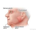
A salivary gland scan uses a special camera and a tracer (radioactive chemical) to take pictures of the salivary glands. This can help your doctor find the cause of dry mouth (xerostomia) or swelling in the salivary glands.
During a salivary gland scan, the tracer liquid is put into a vein (I.V.) in your arm. The tracer moves through your blood and into the salivary glands. A special camera takes pictures to show how much tracer stays in the salivary glands.
Why It Is Done
Why It Is Done
A salivary gland scan is done to:
- Find the cause of swelling in the major salivary glands. Swelling may be caused by an infection (abscess), inflammation, or a pocket of fluid (cyst).
- See if a growth in the parotid gland is a benign tumor or may be cancer.
- Find the cause of dry mouth (xerostomia). Several problems can cause dry mouth, such as a blocked salivary duct, a growth in a salivary gland, or Sjögren's syndrome.
How To Prepare
How To Prepare
Tell your doctor if you are or may be pregnant. If you are breastfeeding, ask your doctor or radiologist if you will need to pause breastfeeding after the test. You may want to pump enough breast milk before the test to get through 1 to 2 days of feeding. Certain radioactive tracers used in this test can get into your breast milk and are not good for the baby.
How It Is Done
How It Is Done
A salivary gland scan is usually done by a nuclear medicine technologist. The pictures are usually interpreted by a radiologist or nuclear medicine specialist.
You will need to take off jewelry that may get in the way of the scan.
During a salivary gland scan, you will sit with the camera placed at your neck. A small amount of the tracer is put in your vein (I.V.).
The camera will scan for radiation released by the tracer. The pictures are taken every few minutes during the scan. You need to stay very still during the scan so the pictures are not blurry.
You may be asked to suck on a lemon after the first pictures are taken. This causes your salivary glands to release more saliva. Then more pictures are taken.
A salivary gland scan takes about 1 hour.
How It Feels
How It Feels
You will not feel pain during the test. You may feel a quick sting or pinch when the I.V. is put in your arm. The tracer may make you feel warm and flushed.
You may find it hard to lie still during the scan.
Risks
Risks
Allergic reactions to the tracer are very rare.
In some cases, soreness or swelling may develop at the I.V. site. Apply a moist, warm compress to your arm to relieve these symptoms.
Anytime you're exposed to radiation, there's a small chance of damage to cells or tissue. That's the case even with the low-level radioactive tracer used for this test. But the chance of damage is very low compared with the benefits of the test.
Most of the tracer will leave your body through your urine or stool within a day. So be sure to flush the toilet right after you use it, and wash your hands well with soap and water. The amount of radiation in the tracer is very small. This means it isn't a risk for people to be around you after the test.
Results
Results
The results of a salivary gland scan are usually available within 2 days.
Normal:
- The tracer moves evenly through the salivary glands and is released normally into the mouth.
- The salivary ducts leading from the salivary glands are not blocked. Saliva is released in response to sucking on a lemon.
Abnormal:
- The tracer does not move evenly through the salivary glands. A pocket of fluid (cyst), a pocket of infection (abscess), or a tumor or other growth may be present.
- The tracer may not flow normally from the salivary glands into the mouth. This may be caused by a tumor pressing on the duct, a stone in the duct, or inflammation of the duct.
- The flow of tracer through the salivary glands is decreased. This may point to a condition such as Sjögren's syndrome.
- The amount of tracer in the salivary glands in front of the ear is greatly increased. This may indicate inflammation or infection of the parotid glands (parotitis).
This information does not replace the advice of a doctor. Ignite Healthwise, LLC, disclaims any warranty or liability for your use of this information. Your use of this information means that you agree to the Terms of Use. Learn how we develop our content.
To learn more about Ignite Healthwise, LLC, visit webmdignite.com.
© 2024-2025 Ignite Healthwise, LLC.
Related Links
Medical Tests: Questions to Ask the Doctor
<cipublic-spinner variant="large"><span>Loading…</span></cipublic-spinner>