Learn about the medical, dental, pharmacy, behavioral, and voluntary benefits your employer may offer.
Liver cancer includes two major types: hepatocellular carcinoma (HCC) and intrahepatic bile duct cancer. For information about bile duct cancer, see Bile Duct Cancer (Cholangiocarcinoma) Treatment. For more information about other, less common types of liver cancer, see the Cellular Classification of Adult Primary Liver Cancer section.
Incidence and Mortality
Estimated new cases and deaths from liver and intrahepatic bile duct cancer in the United States in 2023:[
HCC is relatively uncommon in the United States, although its incidence is rising, principally in relation to the spread of hepatitis C virus infection.[
Anatomy
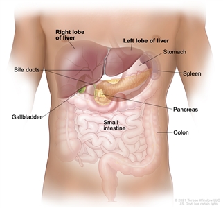
Anatomy of the liver. The liver is in the upper abdomen near the stomach, intestines, gallbladder, and pancreas. The liver has a right lobe and a left lobe. Each lobe is divided into two sections (not shown).
Risk Factors
Increasing age is the most important risk factor for most cancers. Other risk factors for liver (hepatocellular) cancer include the following:
For more information, see Liver (Hepatocellular) Cancer Prevention.
Screening
For more information, see Liver (Hepatocellular) Cancer Screening.
Diagnostic Factors
For lesions that are smaller than 1 cm and are detected during screening in patients at high risk for HCC, further diagnostic evaluation is not required because most of these lesions will be cirrhotic lesions rather than HCC.[
For patients with liver lesions larger than 1 cm who are at risk for HCC, a diagnosis can be considered. The tests required to diagnose HCC may include imaging, biopsy, or both.
Diagnostic imaging
In patients with cirrhosis, liver disease, or other risk factors for HCC, and with lesions greater than 1 cm, triple-phase, contrast-enhanced studies (dynamic computed tomography [CT] or magnetic resonance imaging [MRI]) can be used to establish a diagnosis of HCC.[
A triple-phase CT or MRI assesses the entire liver in distinct phases of perfusion. Following the controlled administration of intravenous contrast media, the arterial and venous phases of perfusion are imaged.
During the arterial phase of the study, HCC enhances more intensely than the surrounding liver because the arterial blood in the liver is diluted by venous blood that does not contain contrast, whereas the HCC contains only arterial blood. In the venous phase, the HCC enhances less than the surrounding liver (which is referred to as the venous washout of HCC), because the arterial blood flowing through the lesion no longer contains contrast; however, the portal blood in the liver now contains contrast.
The presence of arterial uptake followed by washout in a single dynamic study is highly specific (95%–100%) for HCC of 1 to 3 cm in diameter and virtually diagnostic of HCC.[
However, if a first imaging modality, such as a contrast-enhanced CT or MRI, is not conclusive, sequential imaging with a different modality can improve sensitivity for HCC detection (from 33% to 41% for either CT or MRI to 76% for both studies when performed sequentially) without a decrease in specificity.[
If, despite the use of two imaging modalities, a lesion larger than 1 cm remains uncharacterized in a patient at high risk for HCC (i.e., with no or only one classic enhancement pattern), a liver biopsy can be considered.[
Liver biopsy
A liver biopsy may be performed when a diagnosis of HCC is not established by a dynamic imaging modality (three-phase CT or MRI) for liver lesions 1 cm or larger in high-risk patients.
Alpha-fetoprotein (AFP) levels
AFP is insufficiently sensitive or specific for use as a diagnostic assay. AFP can be elevated in intrahepatic cholangiocarcinoma and in some cases in which there are metastases from colon cancer. Finding a mass in the liver of a patient with an elevated AFP does not automatically indicate HCC. However, if the AFP level is high, it can be used to monitor for recurrence.
Prognosis
The natural course of early tumors is poorly known because most HCC patients are treated. However, older reports have described 3-year survival rates of 13% to 21% without any specific treatment.[
Liver transplantation, surgical resection, and ablation offer high rates of complete responses and a potential for cure in patients with early HCC.[
The natural course of advanced-stage HCC is better known. Untreated patients with advanced disease usually survive less than 6 months.[
Unlike most patients with solid tumors, the prognosis of patients with HCC is affected by the tumor stage at presentation and by the underlying liver function. The following prognostic factors guide the selection of treatment:
References:
Malignant primary tumors of the liver consist of two major cell types, which are hepatocellular (90% of cases) and cholangiocarcinoma.[
Histological classification is as follows:
It is important to distinguish between the fibrolamellar variant of HCC and HCC itself because an increased proportion of patients with the fibrolamellar variant may be cured if the tumor can be resected. This variant is found more frequently in young women. It also generally exhibits a slower clinical course than the more common HCC.[
References:
Prognostic modeling in hepatocellular carcinoma (HCC) is complex because cirrhosis is involved in as many as 80% of the cases. Tumor features and the factors related to functional hepatic reserve must be considered. The key prognostic factors are only partially known and vary at different stages of the disease.
More than ten classifications are used throughout the world, but no system is accepted worldwide. New classifications have been proposed to overcome the difficulties of having several staging systems.
This summary discusses the following three staging systems:
Barcelona Clinic Liver Cancer (BCLC) Staging System
Currently, the BCLC staging classification is the most accepted staging system for HCC and is useful in the staging of early tumors. Evidence from an American cohort has shown that BCLC staging offers better prognostic stratification power than other staging systems.[
The BCLC staging system attempts to overcome the limitations of previous staging systems by including variables related to the following:[
Five stages (0 and A through D) are identified based on the variables mentioned above. The BCLC staging system links each HCC stage to appropriate treatment modalities as follows:
Okuda Staging System
The Okuda staging system has been extensively used in the past and includes variables related to tumor burden and liver function, such as bilirubin, albumin, and ascites. However, many significant prognostic tumor factors confirmed in both surgical and nonsurgical series (e.g., unifocal or multifocal, vascular invasion, portal venous thrombosis, or locoregional lymph node involvement) are not included.[
AJCC Staging System and Definitions of TNM
The TNM (tumor, node, metastasis) classification for staging, proposed by the AJCC, is not widely used for liver cancer. Clinical use of TNM staging is limited because liver function is not considered. It is also difficult to use this system to select treatment options because TNM staging relies on detailed histopathological examination available only after tumor excision. TNM may be useful in prognostic prediction after liver resection.[
Stage | TNM | Description |
---|---|---|
Tumor = primary tumor; N = regional lymph nodes; M = distant metastasis. | ||
a Reprinted with permission from AJCC: Liver. In: Amin MB, Edge SB, Greene FL, et al., eds.:AJCC Cancer Staging Manual. 8th ed. New York, NY: Springer, 2017, pp. 287–93. | ||
IA | T1a, N0, M0 | T1a = Solitary tumor ≤2 cm. |
N0 = No regional lymph node metastasis. | ||
M0 = No distant metastasis. | ||
IB | T1b, N0, M0 | T1b = Solitary tumor >2 cm without vascular invasion. |
N0 = No regional lymph node metastasis. | ||
M0 = No distant metastasis. |
Stage | TNM | Description |
---|---|---|
T = primary tumor; N = regional lymph nodes; M = distant metastasis. | ||
a Reprinted with permission from AJCC: Liver. In: Amin MB, Edge SB, Greene FL, et al., eds.:AJCC Cancer Staging Manual. 8th ed. New York, NY: Springer, 2017, pp. 287–93. | ||
II | T2, N0, M0 | T2 = Solitary tumor >2 cm with vascular invasion, or multiple tumors, none >5 cm. |
N0 = No regional lymph node metastasis. | ||
M0 = No distant metastasis. |
Stage | TNM | Description |
---|---|---|
T = primary tumor; N = regional lymph nodes; M = distant metastasis. | ||
a Reprinted with permission from AJCC: Liver. In: Amin MB, Edge SB, Greene FL, et al., eds.:AJCC Cancer Staging Manual. 8th ed. New York, NY: Springer, 2017, pp. 287–93. | ||
IIIA | T3, N0, M0 | T3 = Multiple tumors, at least one of which is >5 cm. |
N0 = No regional lymph node metastasis. | ||
M0 = No distant metastasis. | ||
IIIB | T4, N0, M0 | T4 = Single tumor or multiple tumors of any size involving a major branch of the portal vein or hepatic vein, or tumor(s) with direct invasion of adjacent organs other than the gallbladder or with perforation of visceral peritoneum. |
N0 = No regional lymph node metastasis. | ||
M0 = No distant metastasis. |
Stage | TNM | Description |
---|---|---|
T = primary tumor; N = regional lymph nodes; M = distant metastasis. | ||
a Reprinted with permission from AJCC: Liver. In: Amin MB, Edge SB, Greene FL, et al., eds.:AJCC Cancer Staging Manual. 8th ed. New York, NY: Springer, 2017, pp. 287–93. | ||
IVA | Any T, N1, M0 | TX = Primary tumor cannot be assessed. |
T0 = No evidence of primary tumor. | ||
T1 = Solitary tumor ≤2 cm, or >2 cm without vascular invasion. | ||
–T1a = Solitary tumor ≤2 cm. | ||
–T1b = Solitary tumor >2 cm without vascular invasion. | ||
T2 = Solitary tumor >2 cm with vascular invasion, or multiple tumors, none >5 cm. | ||
T3 = Multiple tumors, at least one of which is >5 cm. | ||
T4 = Single tumor or multiple tumors of any size involving a major branch of the portal vein or hepatic vein, or tumor(s) with direct invasion of adjacent organs other than the gallbladder or with perforation of visceral peritoneum. | ||
N1 = Regional lymph node metastasis. | ||
M0 = No distant metastasis. | ||
IVB | Any T, Any N, M1 | Any T = See descriptions above in this table, stage IVA, Any T, N1, M0. |
NX = Regional lymph nodes cannot be assessed. | ||
N0 = No regional lymph node metastasis. | ||
N1 = Regional lymph node metastasis. | ||
M1 = Distant metastasis. |
References:
There is no agreement on a single treatment strategy for patients with hepatocellular carcinoma (HCC). Selection of treatment is complex due to several factors, including:
Several treatments for HCC are associated with long-term survival, including surgical resection, liver transplantation, and ablation. There are no large, robust, randomized studies that compare treatments considered effective for early-stage disease, nor are there studies comparing these treatments with best supportive care. Often, patients with HCC are evaluated by a multidisciplinary team including hepatologists, radiologists, interventional radiologists, radiation oncologists, transplant surgeons, surgical oncologists, pathologists, and medical oncologists.
Best survivals are achieved when the HCC can be removed either by surgical resection or liver transplantation. Surgical resection is usually performed in patients with localized HCC and enough functional hepatic reserve.
For patients with decompensated cirrhosis and a solitary lesion (<5 cm) or early multifocal disease (≤3 lesions, ≤3 cm in diameter), the best option is liver transplantation,[
Among noncurative treatments for HCC, transarterial chemoembolization and multikinase inhibitors have been shown to improve survival.[
Table 5 shows the standard treatment options for HCC.
Stage | Standard Treatment Options |
---|---|
Localized | Surgical resection |
Liver transplantation | |
Ablation | |
Locally advanced or metastatic | Transarterial embolization and transcatheter arterial chemoembolization |
Targeted therapy (multikinase inhibitors) | |
Second-line immunotherapy | |
Radiation therapy | |
Recurrent Stage | Liver transplantation |
Surgical resection | |
Ablation | |
Palliative therapy |
References:
Localized hepatocellular carcinomas (HCCs) that present as a solitary mass in a portion of the liver or as a limited number of tumors (≤3 lesions, ≤3 cm in diameter) without major vascular invasion constitute approximately 30% of the HCC cases.
There are three potentially curative therapies that are acceptable treatment options for small, single-lesion HCC in patients with well-preserved liver function.
Standard Treatment Options for Localized Adult Primary Liver Cancer
Standard treatment options for localized adult primary liver cancer include the following:
Resection and transplantation achieve the best outcomes in well-selected candidates and are usually considered to be the first option for curative intent.
Surgical resection
Surgery is the mainstay of HCC treatment.
Preoperative assessment includes three-phase helical computed tomography, magnetic resonance imaging, or both to determine the presence of an extension of a tumor across interlobar planes and potential involvement of the hepatic hilus, hepatic veins, and inferior vena cava. Tumors can be resected only if enough liver parenchyma can be spared with adequate vascular and biliary inflow and outflow. Patients with well-compensated cirrhosis can generally tolerate resection of up to 50% of their liver parenchyma.
Surgical resection can be considered for patients who meet the following criteria:
After considering the location and number of tumors, and the hepatic function of the patient, only 5% to 10% of patients with liver cancer will prove to have localized disease amenable to resection.[
The principles of surgical resection involve obtaining a clear margin around the tumor, which may require any of the following:
The 5-year overall survival (OS) rate after curative resection ranges between 27% and 70% and depends on tumor stage and underlying liver function.[
In patients with limited multifocal disease, hepatic resection is controversial.
Liver transplantation
Liver transplantation is a potentially curative therapy for HCC and has the benefit of treating the underlying cirrhosis, but the scarcity of organ donors limits the availability of this treatment modality.[
According to the Milan criteria, patients with a single HCC lesion smaller than 5 cm, or 2 to 3 lesions smaller than 3 cm are eligible for liver transplantation. Expansion of the accepted transplantation criteria for HCC is not supported by consistent data. Liver transplantation is considered if resection is precluded because of multiple, small, tumor lesions (≤3 lesions, each ≤3 cm), or if the liver function is impaired (Child-Pugh class B and class C). In patients who meet the criteria, transplantation is associated with a 5-year OS rate of approximately 70%.[
Ablation
When tumor excision, either by transplant or resection, is not feasible or advisable, ablation may be used if the tumor can be accessed percutaneously or, if necessary, through minimally invasive or open surgery. Ablation may be particularly useful for patients with early-stage HCC that is centrally located in the liver and cannot be surgically removed without excessive sacrifice of functional parenchyma.
Ablation can be achieved in the following ways:
With ablation, a margin of normal liver around the tumor can be considered. Ablation is relatively contraindicated for lesions near bile ducts, the diaphragm, or other intra-abdominal organs that might be injured during the procedure. Furthermore, when tumors are located adjacent to major vessels, the blood flow in the vessels may keep thermal ablation techniques, such as RFA, from reaching optimal temperatures. This is known as the heat-sink effect, which may preclude complete tumor necrosis.
RFA achieves best results in patients with tumors smaller than 3 cm. In this subpopulation of patients, 5-year OS rates may be as high as 59%, and the recurrence-free survival rates may not differ significantly from treatment with hepatic resection.[
PEI obtains good results in patients with Child-Pugh class A cirrhosis and a single tumor smaller than 3 cm in diameter. In those cases, the 5-year OS rate is expected to be as high as 40% to 59%.[
In the few randomized, controlled trials that included patients with Child-Pugh class A cirrhosis, RFA proved superior to PEI in rates of complete response and local recurrences; some of those studies have also shown improved OS with RFA. Furthermore, RFA requires fewer treatment sessions than PEI to achieve comparable outcomes.[
Of note, RFA may have higher complication rates than PEI,[
Treatment Options Under Clinical Evaluation for Localized Adult Primary Liver Cancer
Treatment options under clinical evaluation for localized adult primary liver cancer include the following:
Current Clinical Trials
Use our advanced clinical trial search to find NCI-supported cancer clinical trials that are now enrolling patients. The search can be narrowed by location of the trial, type of treatment, name of the drug, and other criteria. General information about clinical trials is also available.
References:
Standard Treatment Options for Locally Advanced or Metastatic Adult Primary Liver Cancer
Standard treatment options for locally advanced or metastatic adult primary liver cancer not amenable to surgical or locoregional interventions include the following:
Transarterial embolization (TAE) and transcatheter arterial chemoembolization (TACE)
TAE is the most widely used primary treatment for hepatocellular carcinoma (HCC) not amenable to curative treatment by excision or ablation. Most of the blood supply to the normal liver parenchyma comes from the portal vein, whereas blood flow to the tumor comes mainly from the hepatic artery. Furthermore, HCC tumors are generally hypervascular compared with the surrounding normal parenchyma. The obstruction of the arterial branch(es) feeding the tumor may reduce the blood flow to the tumor and result in tumor ischemia and necrosis.
Embolization agents, such as microspheres and particles, may also be administered along with concentrated doses of chemotherapeutic agents (generally doxorubicin or cisplatin) mixed with lipiodol or other emulsifying agents during chemoembolization, arterial chemoembolization (usually via percutaneous access), and TACE. TAE-TACE is considered for patients with HCC who are not amenable to surgery or percutaneous ablation in the absence of extrahepatic disease.
In patients with cirrhosis, any interference with arterial blood supply may be associated with significant morbidity and is relatively contraindicated in the presence of portal hypertension, portal vein thrombosis, or clinical jaundice. In patients with liver decompensation, TAE-TACE could increase the risk of liver failure.
A number of randomized, controlled trials have compared TAE and TACE with supportive care.[
The use of drug-eluting beads (DEB) for TACE has the potential of reducing systemic side effects of chemotherapy and may increase objective tumor response.[
Targeted therapy (multikinase inhibitors)
The U.S. Food and Drug Administration (FDA) has approved two oral multikinase inhibitors, sorafenib and lenvatinib, for first-line treatment of patients with advanced HCC with well-compensated liver function who are not amenable to local therapies. Regorafenib and cabozantinib are approved for second-line treatment of patients with advanced HCC who have progressed on sorafenib.
There are limited data on treatment options for patients with decompensated liver function.
First-line sorafenib
Evidence (sorafenib):
Adverse events attributed to sorafenib in both of these trials included hand-foot skin reaction and diarrhea.[
Studies are also ongoing to evaluate the role of sorafenib after TACE, with chemotherapy, or in the presence of more-advanced liver disease.
First-line lenvatinib
Evidence (lenvatinib):
Second-line regorafenib
Evidence (regorafenib):
Second-line cabozantinib
Evidence (cabozantinib):
While these findings are statistically significant for OS and PFS, the absolute improvement to OS was small, toxicity (including financial toxicity) was high, and the quality-of-life data are lacking to help guide selection of regimen and who should get any treatment. These factors should all be considered and individualized for each patient.
Second-line immunotherapy
Checkpoint inhibitors, particularly programmed death 1 (PD-1) inhibitors are actively being evaluated in the management of advanced HCC.
Nivolumab
Evidence (nivolumab):
Pembrolizumab
Evidence (pembrolizumab):
On the basis of these data, the FDA has granted accelerated approval for pembrolizumab for patients with advanced HCC previously treated with sorafenib.
Radiation therapy
The role of radiation therapy for HCC has traditionally been limited by the low dose tolerance of the liver to radiation. However, recent technological developments in radiation therapy, including breathing-motion management and image-guided radiation therapy, have allowed for more precise and targeted radiation therapy delivery to the liver. Because of these advances, conformal liver irradiation has become feasible in the treatment of focal HCC.
Several phase II studies have suggested a benefit of radiation therapy in local control and OS compared with historical controls for patients with locally advanced HCC unsuitable for standard locoregional therapies.[
Systemic chemotherapy
There is no evidence supporting a survival benefit for patients with advanced HCC receiving systemic cytotoxic chemotherapy when compared with no treatment or best supportive care.
Treatment Options Under Clinical Evaluation for Locally Advanced or Metastatic Adult Primary Liver Cancer
The efficacy of other targeted therapy agents (e.g., sunitinib and brivanib), other checkpoint inhibitors, and combinations of immunotherapy and targeted therapy is being evaluated.
Current Clinical Trials
Use our advanced clinical trial search to find NCI-supported cancer clinical trials that are now enrolling patients. The search can be narrowed by location of the trial, type of treatment, name of the drug, and other criteria. General information about clinical trials is also available.
References:
Treatment Options for Recurrent Adult Primary Liver Cancer
Intrahepatic recurrence is the most common pattern of failure after curative treatment.[
Treatment options for recurrent adult primary liver cancer include the following:
Regarding primary HCC, the treatment strategy for recurrent intrahepatic HCC is determined by the function of the liver and the macroscopic tumor features (e.g., number of lesions, site of recurrence, and invasion of major vessels). Using the same selection criteria that are used for primary HCC, either curative (i.e., salvage liver transplant, surgical resection, and ablation) or palliative treatments (e.g., TACE and sorafenib) can be offered for recurrent HCC.
Evidence (salvage liver transplant, resection, and ablation):
Other studies have also suggested that most of the recurrences that appear early during follow-up are caused by tumor dissemination and have a more aggressive biological pattern than primary tumors.[
Clinical trials are appropriate and can be offered to patients with recurrent HCC whenever possible.
Current Clinical Trials
Use our advanced clinical trial search to find NCI-supported cancer clinical trials that are now enrolling patients. The search can be narrowed by location of the trial, type of treatment, name of the drug, and other criteria. General information about clinical trials is also available.
References:
The PDQ cancer information summaries are reviewed regularly and updated as new information becomes available. This section describes the latest changes made to this summary as of the date above.
General Information About Adult Primary Liver Cancer
Updated statistics with estimated new cases and deaths for 2023 (cited American Cancer Society as reference 1).
This summary is written and maintained by the PDQ Adult Treatment Editorial Board, which is editorially independent of NCI. The summary reflects an independent review of the literature and does not represent a policy statement of NCI or NIH. More information about summary policies and the role of the PDQ Editorial Boards in maintaining the PDQ summaries can be found on the About This PDQ Summary and PDQ® Cancer Information for Health Professionals pages.
Purpose of This Summary
This PDQ cancer information summary for health professionals provides comprehensive, peer-reviewed, evidence-based information about the treatment of adult primary liver cancer. It is intended as a resource to inform and assist clinicians in the care of their patients. It does not provide formal guidelines or recommendations for making health care decisions.
Reviewers and Updates
This summary is reviewed regularly and updated as necessary by the PDQ Adult Treatment Editorial Board, which is editorially independent of the National Cancer Institute (NCI). The summary reflects an independent review of the literature and does not represent a policy statement of NCI or the National Institutes of Health (NIH).
Board members review recently published articles each month to determine whether an article should:
Changes to the summaries are made through a consensus process in which Board members evaluate the strength of the evidence in the published articles and determine how the article should be included in the summary.
The lead reviewers for Adult Primary Liver Cancer Treatment are:
Any comments or questions about the summary content should be submitted to Cancer.gov through the NCI website's Email Us. Do not contact the individual Board Members with questions or comments about the summaries. Board members will not respond to individual inquiries.
Levels of Evidence
Some of the reference citations in this summary are accompanied by a level-of-evidence designation. These designations are intended to help readers assess the strength of the evidence supporting the use of specific interventions or approaches. The PDQ Adult Treatment Editorial Board uses a formal evidence ranking system in developing its level-of-evidence designations.
Permission to Use This Summary
PDQ is a registered trademark. Although the content of PDQ documents can be used freely as text, it cannot be identified as an NCI PDQ cancer information summary unless it is presented in its entirety and is regularly updated. However, an author would be permitted to write a sentence such as "NCI's PDQ cancer information summary about breast cancer prevention states the risks succinctly: [include excerpt from the summary]."
The preferred citation for this PDQ summary is:
PDQ® Adult Treatment Editorial Board. PDQ Adult Primary Liver Cancer Treatment. Bethesda, MD: National Cancer Institute. Updated <MM/DD/YYYY>. Available at: https://www.cancer.gov/types/liver/hp/adult-liver-treatment-pdq. Accessed <MM/DD/YYYY>. [PMID: 26389465]
Images in this summary are used with permission of the author(s), artist, and/or publisher for use within the PDQ summaries only. Permission to use images outside the context of PDQ information must be obtained from the owner(s) and cannot be granted by the National Cancer Institute. Information about using the illustrations in this summary, along with many other cancer-related images, is available in Visuals Online, a collection of over 2,000 scientific images.
Disclaimer
Based on the strength of the available evidence, treatment options may be described as either "standard" or "under clinical evaluation." These classifications should not be used as a basis for insurance reimbursement determinations. More information on insurance coverage is available on Cancer.gov on the Managing Cancer Care page.
Contact Us
More information about contacting us or receiving help with the Cancer.gov website can be found on our Contact Us for Help page. Questions can also be submitted to Cancer.gov through the website's Email Us.
Last Revised: 2023-01-20
This information does not replace the advice of a doctor. Healthwise, Incorporated, disclaims any warranty or liability for your use of this information. Your use of this information means that you agree to the
Healthwise, Healthwise for every health decision, and the Healthwise logo are trademarks of Healthwise, Incorporated.
Individual and family medical and dental insurance plans are insured by Cigna Health and Life Insurance Company (CHLIC), Cigna HealthCare of Arizona, Inc., Cigna HealthCare of Illinois, Inc., Cigna HealthCare of Georgia, Inc., Cigna HealthCare of North Carolina, Inc., Cigna HealthCare of South Carolina, Inc., and Cigna HealthCare of Texas, Inc. Group health insurance and health benefit plans are insured or administered by CHLIC, Connecticut General Life Insurance Company (CGLIC), or their affiliates (see
All insurance policies and group benefit plans contain exclusions and limitations. For availability, costs and complete details of coverage, contact a licensed agent or Cigna sales representative. This website is not intended for residents of New Mexico.