Treatment of Newly Diagnosed Childhood Craniopharyngioma
There is no consensus on the optimal treatment for patients with newly diagnosed craniopharyngioma, in part because of the lack of prospective randomized trials that compare the different treatment options. Treatment is individualized on the basis of the following factors:
- Tumor size.
- Tumor location.
- Extension of the tumor.
- Potential short-term and long-term toxicity, partially related to baseline neuroendocrine and vision deficits (i.e., more conservative surgical approaches may be prioritized in patients who do not have existing neuroendocrine or visual deficits to mitigate the risk of surgical morbidity).[1]
Established treatment options for newly diagnosed childhood craniopharyngioma include the following:
- Complete resection with or without radiation therapy.
- Subtotal resection with radiation therapy.
- Primary cyst drainage with or without radiation therapy.
- Intracystic therapy.
Complete Resection With or Without Radiation Therapy
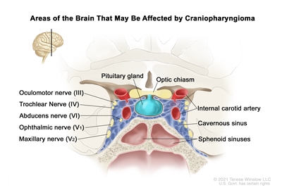
It may be possible to remove all visible tumor and achieve long-term disease control.[2,3,4][Level of evidence C1] A 5-year progression-free survival (PFS) rate of about 65% has been reported.[5] Reported recurrence rates range from less than 10% to nearly 50%.[6,7] Gross-total resection is often technically challenging because the tumor is surrounded by vital structures, including the optic nerves and chiasm, the carotid artery and its branches, the pituitary and hypothalamus, and the third cranial nerve. These structures may limit the ability to remove the entire tumor. Conservative surgical approaches are often used to preserve functional and quality-of-life outcomes.[8,9][Level of evidence C1]
Many surgical approaches have been described, and the choice is determined by tumor size, location, extension, and the patient's baseline signs and symptoms of disease. Surgical approaches include the following:
- Craniotomy: As noted above, gross-total resection may be technically challenging because the tumor is surrounded by vital structures. The surgeon often has a limited view of the hypothalamic and sellar regions, and portions of the mass may remain after surgery, accounting for some recurrences. An understanding of the complex variations in how the tumors grow anatomically may help facilitate gross-total resection.[10] Nonetheless, almost all craniopharyngiomas attach to the pituitary stalk. Of the patients who undergo complete resection, virtually all will require lifelong pituitary hormone replacement with multiple medications.[3,11]
- Transsphenoidal approach: A transsphenoidal approach has been proven possible in patients of all ages and for tumors of various sizes localized within the sella.[12]; [13][Level of evidence C1] The development of expanded endonasal techniques with endoscopic visualization has allowed increased use of this approach, even for sizeable childhood tumors, which is similar to the experience in adults.[14] A complete resection can be obtained using this approach, with associated complications of panhypopituitarism and the risk of cerebrospinal fluid leaks.[15,16] When an endonasal approach is not possible, a craniotomy is required.
Complications of complete resection using either approach include the following:
- Obesity, which can be life-threatening.[17]
- Need for hormone replacement therapy.[18]
- Severe behavioral problems.[18]
- Blindness.
- Seizures.
- Spinal fluid leak.
- False aneurysms.
- Difficulty with eye movements.
- Death from intraoperative hemorrhage, hypothalamic damage, or stroke (rare).
If the surgeon indicates that the tumor was not completely removed or if postoperative imaging reveals residual craniopharyngioma, radiation therapy may be recommended to prevent early progression.[19][Level of evidence C2] For more information, see the Subtotal Resection With Radiation Therapy section.
Routine surveillance using magnetic resonance imaging is performed for several years after complete resection because of the possibility of tumor recurrence.
Subtotal Resection With Radiation Therapy
The goal of limited surgery can be to establish a diagnosis, drain cystic components of the tumor, and decompress surrounding anatomical structures. In subtotal resections, removal of the tumor from the pituitary stalk or hypothalamus is typically avoided to minimize the late effects associated with complete resection.[20]
Surgery is often followed by radiation therapy, because radiation therapy can decrease the risk of recurrence after a subtotal resection.[21] With this approach, the 5-year PFS rates are approximately 70% to 90%,[5,22,23,24,25]; [26][Level of evidence C1] and the 10-year overall survival (OS) rates exceed 90%, which are similar to the rates in patients who undergo a gross-total resection.[27,28][Level of evidence C1]; [29][Level of evidence C2] Most often, radiation therapy is timed to immediately follow subtotal resection. However, in certain cases, such as in young patients or in patients without existing neuroendocrine or visual deficits, serial imaging may be used to delay or avoid radiation therapy for as long as feasible.[7,30] The standard approach to radiation therapy involves fractionated external-beam radiation, with a recommended dose of 50 to 54 Gy, in 1.8-Gy fractions, restricting the optic chiasm dose to 54 Gy.[31,32,33,34] Newer radiation technologies such as intensity-modulated photon therapy and proton-beam radiation therapy may reduce the radiation dose to uninvolved parts of the brain and spare normal tissue.[23,34,35,36] It is unknown whether such techniques reduce the late effects of radiation therapy.[26,34,36,37] Transient cyst enlargement may be noted during radiation therapy, and serial imaging may be required during radiation therapy to assess cyst changes and consider updates to radiation mapping.[38][Level of evidence C3]
Surgical complications with a subtotal resection can be similar to, but are less likely than, with a complete resection. If radiation therapy is used, additional complications must be considered, including the following:
- Loss of pituitary hormonal function.
- Cognitive dysfunction.
- Development of late strokes and vascular malformations.
- Delayed blindness.
- Development of second tumors.
- Malignant transformation of the primary tumor within the radiation field (rare).[39,40]
A phase II single-arm study included 94 patients (aged 12 months to 21 years) with craniopharyngiomas who were treated with proton-beam radiation therapy after individualized surgical resection. These patients were compared with a historical cohort of patients who were treated with photon-beam radiation therapy.[41] The survival outcomes of patients who received proton therapy were similar to those of patients who received photon therapy. The cumulative incidence rates of necrosis, vasculopathy, changes in vision, and severe complications were also similar between the two groups of patients. However, patients treated with proton therapy in the more recent cohort had superior cognitive outcomes.
A long-term study of 101 children who were treated for craniopharyngiomas evaluated visual, neurocognitive, and endocrine outcomes after photon radiation therapy. Race and presence of a shunt affected baseline scores.[42] For children who presented with lower intelligence quotient (IQ) scores at diagnosis, the impact of treatment often resulted in an IQ score reduction to the borderline mental disability range of 70 to 84. The investigators demonstrated that age at treatment (younger children had worse outcomes), radiation dose to the temporal lobes and hippocampi, and visual impairment significantly impacted neurocognitive function after radiation therapy. This study demonstrates the importance of these factors in the treatment and late effects of craniopharyngioma.
A report from the prospective registry study KiProReg examined the use of proton-beam therapy in 84 children younger than 18 years with craniopharyngioma.[43] The estimated 3-year OS rate was 98.2%, and the PFS rate was 94.7%. With a median follow-up of 4.3 years, late toxicities appeared acceptable. Sixty-three of the patients were treated with pencil-beam scanning, which is considered an advancement in proton technology.
Primary Cyst Drainage With or Without Radiation Therapy
For predominantly cystic craniopharyngiomas, stereotactic drainage of the cyst, insertion of a catheter from which drainage can be facilitated, or cyst fenestration are other therapeutic alternatives.[7,44] This can be followed by observation or radiation therapy, depending on clinical and tumor characteristics . This procedure may also allow the surgeon to use the following two-staged approach:[45]
- Draining the cyst to relieve pressure and complicating symptoms.
- Resecting the tumor or employing radiation therapy later.
Intracystic Therapy
Intracystic therapies include peginterferon alpha, radioactive phosphorus P 32 (32P) or other compounds,[46,47,48]; [49][Level of evidence B4] and interferon-alpha (which is no longer commercially available).[50]; [51][Level of evidence C1]; [52][Level of evidence C2] Bleomycin has previously been used.[53]; [54][Level of evidence C2]
A systematic review of publications on the treatment of cystic craniopharyngiomas with radioisotope brachytherapy from 2010 to 2021 identified 66 pediatric patients (N = 228).[55] With a minimum follow-up of 5 years, partial and complete responses were achieved in 89% of children with purely cystic lesions, compared with 58% of children with nonexclusively cystic lesions. Visual improvement was achieved in 64% of the patients with purely cystic lesions, and endocrine improvement was achieved in 20% of these patients. The observed progression rate was 3% for patients with purely cystic lesions. Treatment with intracystic brachytherapy, most commonly using 32P and yttrium Y 90, can be considered for patients with purely cystic craniopharyngiomas.
Treatment Options Under Clinical Evaluation
Information about NCI-supported clinical trials can be found on the NCI website. For information about clinical trials sponsored by other organizations, see the ClinicalTrials.gov website.
Preclinical contemporary evaluations have identified active molecular and immune pathways in craniopharyngioma that may be targetable using commercially available or investigational agents. Specifically, MAPK and RAF pathways and immune/inflammatory targets such as PD-1 pathway components and IL-6 have been identified.[7,56,57,58,59,60,61][Level of evidence C1]
The following is an example of a national and/or institutional clinical trial that is currently being conducted:
- NCT05465174 (Nivolumab and Tovorafenib [DAY101] for Treatment of Craniopharyngioma in Children and Young Adults): This study assesses the tolerability and efficacy of combination therapy with PD-1 (nivolumab) and pan-RAF kinase (tovorafenib) inhibition for the treatment of children and young adults with craniopharyngioma.
References:
- Cohen M, Bartels U, Branson H, et al.: Trends in treatment and outcomes of pediatric craniopharyngioma, 1975-2011. Neuro Oncol 15 (6): 767-74, 2013.
- Mortini P, Losa M, Pozzobon G, et al.: Neurosurgical treatment of craniopharyngioma in adults and children: early and long-term results in a large case series. J Neurosurg 114 (5): 1350-9, 2011.
- Elliott RE, Hsieh K, Hochm T, et al.: Efficacy and safety of radical resection of primary and recurrent craniopharyngiomas in 86 children. J Neurosurg Pediatr 5 (1): 30-48, 2010.
- Zhang YQ, Ma ZY, Wu ZB, et al.: Radical resection of 202 pediatric craniopharyngiomas with special reference to the surgical approaches and hypothalamic protection. Pediatr Neurosurg 44 (6): 435-43, 2008.
- Yang I, Sughrue ME, Rutkowski MJ, et al.: Craniopharyngioma: a comparison of tumor control with various treatment strategies. Neurosurg Focus 28 (4): E5, 2010.
- Müller HL, Merchant TE, Puget S, et al.: New outlook on the diagnosis, treatment and follow-up of childhood-onset craniopharyngioma. Nat Rev Endocrinol 13 (5): 299-312, 2017.
- Apps JR, Muller HL, Hankinson TC, et al.: Contemporary Biological Insights and Clinical Management of Craniopharyngioma. Endocr Rev 44 (3): 518-538, 2023.
- Lohkamp LN, Kasper EM, Pousa AE, et al.: An update on multimodal management of craniopharyngioma in children. Front Oncol 13: 1149428, 2023.
- Bogusz A, Müller HL: Childhood-onset craniopharyngioma: latest insights into pathology, diagnostics, treatment, and follow-up. Expert Rev Neurother 18 (10): 793-806, 2018.
- Morisako H, Goto T, Goto H, et al.: Aggressive surgery based on an anatomical subclassification of craniopharyngiomas. Neurosurg Focus 41 (6): E10, 2016.
- Sands SA, Milner JS, Goldberg J, et al.: Quality of life and behavioral follow-up study of pediatric survivors of craniopharyngioma. J Neurosurg 103 (4 Suppl): 302-11, 2005.
- Bakhsheshian J, Jin DL, Chang KE, et al.: Risk factors associated with the surgical management of craniopharyngiomas in pediatric patients: analysis of 1961 patients from a national registry database. Neurosurg Focus 41 (6): E8, 2016.
- Locatelli D, Massimi L, Rigante M, et al.: Endoscopic endonasal transsphenoidal surgery for sellar tumors in children. Int J Pediatr Otorhinolaryngol 74 (11): 1298-302, 2010.
- Chivukula S, Koutourousiou M, Snyderman CH, et al.: Endoscopic endonasal skull base surgery in the pediatric population. J Neurosurg Pediatr 11 (3): 227-41, 2013.
- Mazzatenta D, Zoli M, Guaraldi F, et al.: Outcome of Endoscopic Endonasal Surgery in Pediatric Craniopharyngiomas. World Neurosurg 134: e277-e288, 2020.
- Lee JA, Cooper RL, Nguyen SA, et al.: Endonasal Endoscopic Surgery for Pediatric Sellar and Suprasellar Lesions: A Systematic Review and Meta-analysis. Otolaryngol Head Neck Surg 163 (2): 284-292, 2020.
- Müller HL, Gebhardt U, Teske C, et al.: Post-operative hypothalamic lesions and obesity in childhood craniopharyngioma: results of the multinational prospective trial KRANIOPHARYNGEOM 2000 after 3-year follow-up. Eur J Endocrinol 165 (1): 17-24, 2011.
- Clark AJ, Cage TA, Aranda D, et al.: Treatment-related morbidity and the management of pediatric craniopharyngioma: a systematic review. J Neurosurg Pediatr 10 (4): 293-301, 2012.
- Lin LL, El Naqa I, Leonard JR, et al.: Long-term outcome in children treated for craniopharyngioma with and without radiotherapy. J Neurosurg Pediatr 1 (2): 126-30, 2008.
- Elowe-Gruau E, Beltrand J, Brauner R, et al.: Childhood craniopharyngioma: hypothalamus-sparing surgery decreases the risk of obesity. J Clin Endocrinol Metab 98 (6): 2376-82, 2013.
- Müller HL: Childhood craniopharyngioma: current controversies on management in diagnostics, treatment and follow-up. Expert Rev Neurother 10 (4): 515-24, 2010.
- Winkfield KM, Tsai HK, Yao X, et al.: Long-term clinical outcomes following treatment of childhood craniopharyngioma. Pediatr Blood Cancer 56 (7): 1120-6, 2011.
- Jimenez RB, Ahmed S, Johnson A, et al.: Proton Radiation Therapy for Pediatric Craniopharyngioma. Int J Radiat Oncol Biol Phys 110 (5): 1480-1487, 2021.
- Eveslage M, Calaminus G, Warmuth-Metz M, et al.: The Postopera tive Quality of Life in Children and Adolescents with Craniopharyngioma. Dtsch Arztebl Int 116 (18): 321-328, 2019.
- Merchant TE, Hua CH, Shukla H, et al.: Proton versus photon radiotherapy for common pediatric brain tumors: comparison of models of dose characteristics and their relationship to cognitive function. Pediatr Blood Cancer 51 (1): 110-7, 2008.
- Merchant TE, Kun LE, Hua CH, et al.: Disease control after reduced volume conformal and intensity modulated radiation therapy for childhood craniopharyngioma. Int J Radiat Oncol Biol Phys 85 (4): e187-92, 2013.
- Schoenfeld A, Pekmezci M, Barnes MJ, et al.: The superiority of conservative resection and adjuvant radiation for craniopharyngiomas. J Neurooncol 108 (1): 133-9, 2012.
- Edmonston DY, Wu S, Li Y, et al.: Limited surgery and conformal photon radiation therapy for pediatric craniopharyngioma: long-term results from the RT1 protocol. Neuro Oncol 24 (12): 2200-2209, 2022.
- Clark AJ, Cage TA, Aranda D, et al.: A systematic review of the results of surgery and radiotherapy on tumor control for pediatric craniopharyngioma. Childs Nerv Syst 29 (2): 231-8, 2013.
- Beckhaus J, Friedrich C, Boekhoff S, et al.: Outcome after pediatric craniopharyngioma: the role of age at diagnosis and hypothalamic damage. Eur J Endocrinol 188 (3): , 2023.
- Kiehna EN, Merchant TE: Radiation therapy for pediatric craniopharyngioma. Neurosurg Focus 28 (4): E10, 2010.
- Harrabi SB, Adeberg S, Welzel T, et al.: Long term results after fractionated stereotactic radiotherapy (FSRT) in patients with craniopharyngioma: maximal tumor control with minimal side effects. Radiat Oncol 9: 203, 2014.
- Lo AC, Howard AF, Nichol A, et al.: Long-term outcomes and complications in patients with craniopharyngioma: the British Columbia Cancer Agency experience. Int J Radiat Oncol Biol Phys 88 (5): 1011-8, 2014.
- Bishop AJ, Greenfield B, Mahajan A, et al.: Proton beam therapy versus conformal photon radiation therapy for childhood craniopharyngioma: multi-institutional analysis of outcomes, cyst dynamics, and toxicity. Int J Radiat Oncol Biol Phys 90 (2): 354-61, 2014.
- Winkfield KM, Linsenmeier C, Yock TI, et al.: Surveillance of craniopharyngioma cyst growth in children treated with proton radiotherapy. Int J Radiat Oncol Biol Phys 73 (3): 716-21, 2009.
- Beltran C, Roca M, Merchant TE: On the benefits and risks of proton therapy in pediatric craniopharyngioma. Int J Radiat Oncol Biol Phys 82 (2): e281-7, 2012.
- Boehling NS, Grosshans DR, Bluett JB, et al.: Dosimetric comparison of three-dimensional conformal proton radiotherapy, intensity-modulated proton therapy, and intensity-modulated radiotherapy for treatment of pediatric craniopharyngiomas. Int J Radiat Oncol Biol Phys 82 (2): 643-52, 2012.
- Shi Z, Esiashvili N, Janss AJ, et al.: Transient enlargement of craniopharyngioma after radiation therapy: pattern of magnetic resonance imaging response following radiation. J Neurooncol 109 (2): 349-55, 2012.
- Ishida M, Hotta M, Tsukamura A, et al.: Malignant transformation in craniopharyngioma after radiation therapy: a case report and review of the literature. Clin Neuropathol 29 (1): 2-8, 2010 Jan-Feb.
- Aquilina K, Merchant TE, Rodriguez-Galindo C, et al.: Malignant transformation of irradiated craniopharyngioma in children: report of 2 cases. J Neurosurg Pediatr 5 (2): 155-61, 2010.
- Merchant TE, Hoehn ME, Khan RB, et al.: Proton therapy and limited surgery for paediatric and adolescent patients with craniopharyngioma (RT2CR): a single-arm, phase 2 study. Lancet Oncol 24 (5): 523-534, 2023.
- Merchant TE, Dangda S, Hoehn ME, et al.: Pediatric Craniopharyngioma: The Effect of Visual Deficits and Hormone Deficiencies on Long-Term Cognitive Outcomes After Conformal Photon Radiation Therapy. Int J Radiat Oncol Biol Phys 115 (3): 581-591, 2023.
- Bischoff M, Khalil DA, Frisch S, et al.: Outcome After Modern Proton Beam Therapy in Childhood Craniopharyngioma: Results of the Prospective Registry Study KiProReg. Int J Radiat Oncol Biol Phys 120 (1): 137-148, 2024.
- Cinalli G, Spennato P, Cianciulli E, et al.: The role of transventricular neuroendoscopy in the management of craniopharyngiomas: three patient reports and review of the literature. J Pediatr Endocrinol Metab 19 (Suppl 1): 341-54, 2006.
- Schubert T, Trippel M, Tacke U, et al.: Neurosurgical treatment strategies in childhood craniopharyngiomas: is less more? Childs Nerv Syst 25 (11): 1419-27, 2009.
- Julow J, Backlund EO, Lányi F, et al.: Long-term results and late complications after intracavitary yttrium-90 colloid irradiation of recurrent cystic craniopharyngiomas. Neurosurgery 61 (2): 288-95; discussion 295-6, 2007.
- Barriger RB, Chang A, Lo SS, et al.: Phosphorus-32 therapy for cystic craniopharyngiomas. Radiother Oncol 98 (2): 207-12, 2011.
- Maarouf M, El Majdoub F, Fuetsch M, et al.: Stereotactic intracavitary brachytherapy with P-32 for cystic craniopharyngiomas in children. Strahlenther Onkol 192 (3): 157-65, 2016.
- Kickingereder P, Maarouf M, El Majdoub F, et al.: Intracavitary brachytherapy using stereotactically applied phosphorus-32 colloid for treatment of cystic craniopharyngiomas in 53 patients. J Neurooncol 109 (2): 365-74, 2012.
- Ierardi DF, Fernandes MJ, Silva IR, et al.: Apoptosis in alpha interferon (IFN-alpha) intratumoral chemotherapy for cystic craniopharyngiomas. Childs Nerv Syst 23 (9): 1041-6, 2007.
- Cavalheiro S, Di Rocco C, Valenzuela S, et al.: Craniopharyngiomas: intratumoral chemotherapy with interferon-alpha: a multicenter preliminary study with 60 cases. Neurosurg Focus 28 (4): E12, 2010.
- Kilday JP, Caldarelli M, Massimi L, et al.: Intracystic interferon-alpha in pediatric craniopharyngioma patients: an international multicenter assessment on behalf of SIOPE and ISPN. Neuro Oncol 19 (10): 1398-1407, 2017.
- Linnert M, Gehl J: Bleomycin treatment of brain tumors: an evaluation. Anticancer Drugs 20 (3): 157-64, 2009.
- Hukin J, Steinbok P, Lafay-Cousin L, et al.: Intracystic bleomycin therapy for craniopharyngioma in children: the Canadian experience. Cancer 109 (10): 2124-31, 2007.
- Guimarães MM, Cardeal DD, Teixeira MJ, et al.: Brachytherapy in paediatric craniopharyngiomas: a systematic review and meta-analysis of recent literature. Childs Nerv Syst 38 (2): 253-262, 2022.
- Petralia F, Tignor N, Reva B, et al.: Integrated Proteogenomic Characterization across Major Histological Types of Pediatric Brain Cancer. Cell 183 (7): 1962-1985.e31, 2020.
- Apps JR, Carreno G, Gonzalez-Meljem JM, et al.: Tumour compartment transcriptomics demonstrates the activation of inflammatory and odontogenic programmes in human adamantinomatous craniopharyngioma and identifies the MAPK/ERK pathway as a novel therapeutic target. Acta Neuropathol 135 (5): 757-777, 2018.
- Hengartner AC, Prince E, Vijmasi T, et al.: Adamantinomatous craniopharyngioma: moving toward targeted therapies. Neurosurg Focus 48 (1): E7, 2020.
- Coy S, Rashid R, Lin JR, et al.: Multiplexed immunofluorescence reveals potential PD-1/PD-L1 pathway vulnerabilities in craniopharyngioma. Neuro Oncol 20 (8): 1101-1112, 2018.
- Grob S, Mirsky DM, Donson AM, et al.: Targeting IL-6 Is a Potential Treatment for Primary Cystic Craniopharyngioma. Front Oncol 9: 791, 2019.
- Donson AM, Apps J, Griesinger AM, et al.: Molecular Analyses Reveal Inflammatory Mediators in the Solid Component and Cyst Fluid of Human Adamantinomatous Craniopharyngioma. J Neuropathol Exp Neurol 76 (9): 779-788, 2017.