Learn about the medical, dental, pharmacy, behavioral, and voluntary benefits your employer may offer.
Retinoblastoma is a pediatric cancer that requires careful integration of multidisciplinary care. Treatment of retinoblastoma aims to save the patient's life and preserve useful vision. For patients presenting with extraocular retinoblastoma, treatment with systemic chemotherapy and radiation therapy is likely to be curative. However, extraorbital disease requires intensive chemotherapy and may include consolidation with high-dose chemotherapy and autologous hematopoietic stem cell rescue with or without radiation therapy. While a large proportion of patients with systemic extra–central nervous system (CNS) metastases can be cured, the prognosis for patients with intracranial disease is dismal.
Incidence
Retinoblastoma is a relatively uncommon tumor of childhood that arises in the retina and accounts for about 3% of the cancers occurring in children younger than 15 years.
Retinoblastoma is a cancer of the very young child; two-thirds of all cases of retinoblastoma are diagnosed before age 2 years.[
Anatomy
Retinoblastoma arises from the retina, and it may grow under the retina and/or toward the vitreous cavity. Involvement of the ocular coats and optic nerve occurs as a sequence of events as the tumor progresses.
Focal invasion of the choroid is common, although massive invasion occurs in cases of advanced disease. After invading the choroid, the tumor gains access to systemic circulation and creates the potential for metastases. Further progression through the ocular coats leads to invasion of the sclera and the orbit. Tumors that invade the anterior chamber may gain access to systemic circulation through the canal of Schlemm. Progression through the optic nerve and past the lamina cribrosa increases the risk of systemic and CNS dissemination (see Figure 1).
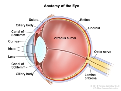
Figure 1. Anatomy of the eye showing the sclera, ciliary body, canal of Schlemm, cornea, iris, lens, vitreous humor, retina, choroid, optic nerve, and lamina cribrosa. The vitreous humor is a gel that fills the center of the eye.
Screening
Consensus reports from the American Association of Ophthalmic Oncologists and Pathologists and the American Association for Cancer Research Childhood Cancer Predisposition Workshop describe surveillance guidelines for screening children at risk of developing retinoblastoma.[
In children with a positive family history of retinoblastoma, early-in-life screening by fundus exam is performed under general anesthesia at regular intervals according to a schedule based on the absolute estimated risk, as determined by the identification of the RB1 mutation in the family and the presence of the RB1 mutation in the child.[
Infants born to affected parents have a dilated eye examination under anesthesia as soon as possible in the first month of life, and a genetic evaluation is performed. Infants with a positive genetic test are examined under anesthesia on a monthly basis. In infants who do not develop disease, monthly exams continue throughout the first year. The frequency of those exams may be decreased progressively during the second and subsequent years. Screening children with a positive family history of retinoblastoma can improve their prognosis, in terms of globe sparing and use of less intensive, ocular-salvage treatments (see Table 1 and Figure 2).[
Relative of Proband | Pretest Risk for Mutant Allele (%) | |
---|---|---|
| Bilateral Proband (100) | Unilateral Proband (15) |
a Reprinted fromOphthalmology, Volume 125, Issue 3, Alison H. Skalet, Dan S. Gombos, Brenda L. Gallie, Jonathan W. Kim, Carol L. Shields, Brian P. Marr, Sharon E. Plon, Patricia Chévez-Barrios, Screening Children at Risk for Retinoblastoma: Consensus Report from the American Association of Ophthalmic Oncologists and Pathologists, Pages 453–458, Copyright (2018), with permission from Elsevier. | ||
b Pretest risk forRB1mutation in family members of an affected child withretinoblastoma. Risk forRB1mutant allele is shown as a percentage for unilateral and bilateral probands without family history of retinoblastoma. | ||
c Third- and fourth-degree relatives of unilateral probands have calculated risks of 0.003% and 0.001%, respectively, which are less than the normal population risk of 0.007% (1 in 15,000 live births); therefore, the risk is stated at 0.007%. | ||
Offspring (infant) | 50 | 7.5 |
Parent | 5 | 0.8 |
Sibling | 2.5 | 0.4 |
Niece/nephew | 1.3 | 0.2 |
Aunt/uncle | 0.1 | 0.007c |
First cousin | 0.05 | 0.007c |
General population | 0.007 |
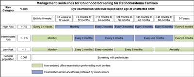
Figure 2. Management guidelines for childhood screening for retinoblastoma. The presented schedules are general guidelines and reflect a schedule for examinations in which no lesions of concern are noted. It may be appropriate to examine some children more frequently. Decisions regarding examination method, examination under anesthesia (EUA) versus nonsedated examination in the office, are complex and best decided by the clinician in discussion with the patient's family. The preference of the majority of the clinical centers involved in the creation of this consensus statement is reflected, but individual centers may make policy decisions based on available resources and expert clinician preference. Examination under anesthesia will be strongly considered for any child who is unable to participate in an office examination sufficiently to allow thorough examination of the retina. *A minority of clinical centers also prefer EUA for high- and intermediate-risk children (calculated risk >1%) from birth to 8 weeks of age. Reprinted from Ophthalmology, Volume 125, Issue 3, Alison H. Skalet, Dan S. Gombos, Brenda L. Gallie, Jonathan W. Kim, Carol L. Shields, Brian P. Marr, Sharon E. Plon, Patricia Chévez-Barrios, Screening Children at Risk for Retinoblastoma: Consensus Report from the American Association of Ophthalmic Oncologists and Pathologists, Pages 453–458, Copyright (2018), with permission from Elsevier.
It is common practice to use ophthalmic examinations to screen the parents and siblings of patients with retinoblastoma to exclude an unknown familial disease. However, in the absence of genetic testing, the screening plan for a child with a biological parent who had unilateral retinoblastoma is not well defined.[
Clinical Presentation
Age at presentation correlates with laterality. Patients with bilateral disease present at a younger age, usually in the first 12 months of life.
Most patients present with leukocoria, which is occasionally first noticed after a flash photograph is taken. Strabismus is the second most common presenting sign and usually correlates with macular involvement. Very advanced intraocular tumors present with pain, orbital cellulitis, glaucoma, or buphthalmos.
As the tumor progresses, patients may present with orbital or metastatic disease. Metastases occur in the preauricular and laterocervical lymph nodes, in the CNS, or systemically (commonly in the bones, bone marrow, and liver).
In the United States, Hispanic children and children living in lower socioeconomic conditions have presented with more advanced disease.[
Diagnostic and Staging Evaluation
Diagnostic evaluation of retinoblastoma includes the following:
Patients with suspected extraocular extension by imaging or high-risk pathology in the enucleated eye (i.e., massive choroidal invasion or involvement of the sclera or the optic nerve beyond the lamina cribrosa) may need to be evaluated for the presence of metastatic disease. Patients presenting with these pathological features in the enucleated eye are at high risk of developing metastases. In these cases, the following procedures may be performed:[
Genetics and Genomics of Retinoblastoma
Genomics of Retinoblastoma
Retinoblastoma is a tumor that occurs in heritable (25%–30%) and nonheritable (70%–75%) forms. Heritable disease is defined by the presence of a germline mutation of the RB1 gene. This germline mutation may have been inherited from an affected progenitor (25% of cases) or may have occurred in a germ cell before conception or in utero during early embryogenesis in patients with sporadic disease (75% of cases). The presence of positive family history or bilateral or multifocal disease is suggestive of heritable disease.
Heritable retinoblastoma may manifest as unilateral or bilateral disease. The penetrance of the RB1 mutation (laterality, age at diagnosis, and number of tumors) is probably dependent on concurrent genetic modifiers such as MDM2 and MDM4 polymorphisms.[
Children with heritable retinoblastoma tend to be diagnosed at a younger age than are children with the nonheritable form of the disease.
The genomic landscape of retinoblastoma is driven by alterations in RB1 that lead to biallelic inactivation.[
Recurrent changes in genes other than RB1 are uncommon in retinoblastoma but do occur. Mutations or deletions of BCOR and amplification of MYCN are the most frequently reported events.[
Genetic counseling is recommended for all patients with retinoblastoma. For more information, see the Genetic Counseling section.
Genetic Testing
Blood and tumor samples can be tested to determine whether a patient with retinoblastoma has a germline or somatic mutation in the RB1 gene. Once the patient's genetic mutation has been identified, other family members can be screened directly for the mutation with targeted sequencing.
A multistep assay that includes the following may be performed for a complete genetic evaluation of the RB1 gene:[
In cases of somatic mosaicism or cytogenetic abnormalities, the mutations may not be easily detected. More exhaustive techniques such as karyotyping, fluorescence in situ hybridization, and methylation analysis of the RB1 promoter may be needed. Deep (2500x) sequencing of an RB1 genomic amplicon from lymphocyte DNA can reveal low-level mosaicism.[
The absence of detectable somatic RB1 mutations in approximately 3% of unilateral, nonheritable retinoblastoma cases suggests that alternative genetic mechanisms may underlie the development of retinoblastoma.[
Genetic Counseling
Genetic counseling is an integral part of the management of patients with retinoblastoma and their families, regardless of clinical presentation. Counseling includes a discussion of the main forms of retinoblastoma, which assists parents in understanding the genetic consequences of each form of retinoblastoma and in estimating the risk of disease in family members.[
Genetic counseling, however, is not always straightforward. Approximately 10% of children with retinoblastoma have somatic genetic mosaicism, which contributes to the difficulty of genetic counseling.[
Postdiagnosis Surveillance
Children with a germline RB1 mutation may continue to develop new tumors for a few years after diagnosis and treatment. For this reason, these patients need to be examined frequently. It is common practice for examinations to occur every 2 to 4 months for at least 28 months.[
A proportion of children who present with unilateral retinoblastoma will eventually develop disease in the opposite eye. Periodic examinations of the unaffected eye are performed until the germline status of the RB1 gene is determined.
Because of the poor prognosis for patients with trilateral retinoblastoma, screening with neuroimaging until age 5 years is a common practice in the monitoring of children with the heritable form of the disease. For more information, see the Trilateral retinoblastoma section.
Causes of Retinoblastoma-Related Mortality
While retinoblastoma is a highly curable disease, the challenge for those who treat retinoblastoma is to preserve life and to prevent the loss of an eye, blindness, and other serious effects of treatment that reduce the patient's life span or quality of life. With improvements in the diagnosis and management of retinoblastoma over the past several decades, metastatic retinoblastoma is observed less frequently in the United States and other developed nations. As a result, other causes, such as trilateral retinoblastoma and subsequent neoplasms (SNs), have become significant contributors to retinoblastoma-related mortality in the first and subsequent decades of life. In the United States, before the advent of chemoreduction as a means of treating heritable or bilateral disease and the implementation of neuroimaging screening, trilateral retinoblastoma contributed to more than 50% of retinoblastoma-related mortality for patients in the first decade after their diagnosis.[
Trilateral retinoblastoma
Trilateral retinoblastoma is a well-recognized syndrome that occurs in 5% to 15% of patients with heritable retinoblastoma. It is defined by the development of an asynchronous intracranial midline neuroblastic tumor, which typically develops between the ages of 20 and 36 months.[
Because of its poor prognosis and the apparent improved survival with early detection and aggressive treatment, screening with routine neuroimaging could potentially detect most cases within 2 years of first diagnosis.[
Although it is not clear whether early diagnosis can impact survival, screening with MRI has been recommended as often as every 6 months for 5 years for patients suspected of having heritable disease or those with unilateral disease and a positive family history.[
A cystic pineal gland, which is commonly detected by surveillance MRI, needs to be distinguished from a cystic variant of pineoblastoma. In children without retinoblastoma, the incidence of pineal cysts has been reported to be 55.8%.[
References:
Maturing cone precursor cells appear to be the cell of origin in human retinoblastoma.[
Retinoblastomas are characterized by marked cell proliferation, as evidenced by high mitosis counts, extremely high MIB-1 labeling indices, and strong diffuse nuclear immunoreactivity for CRX, a useful marker to discriminate retinoblastoma from other malignant, small, round cell tumors.[
Cavitary retinoblastoma, a rare variant of retinoblastoma, has ophthalmoscopically visible lucent cavities within the tumor. The cavitary spaces appear hollow on ultrasonography and hypofluorescent on angiography. Histopathologically, the cavitary spaces have been shown to represent areas of photoreceptor differentiation.[
Cavitary retinoblastoma has been associated with minimal visible response to intravenous and intra-arterial chemotherapy, which is thought to be a sign of tumor differentiation.[
A pathologist experienced in ocular pathology and retinoblastoma should examine the enucleated specimen, particularly to determine risk features of extraocular dissemination. For more information, see the Treatment of Intraocular Retinoblastoma section.
References:
The staging of patients with retinoblastoma requires close coordination of radiologists, pediatric oncologists, and ophthalmologists. Several staging and grouping systems have been proposed for retinoblastoma.[
Intraocular Retinoblastoma
Intraocular retinoblastoma is localized to the eye. It may be confined to the retina or may extend to involve other structures such as the choroid, ciliary body, anterior chamber, and optic nerve head. Intraocular retinoblastoma, however, does not extend beyond the eye into the tissues around the eye or to other parts of the body.
Extraocular Retinoblastoma
Extraocular retinoblastoma extends beyond the eye. It may be confined to the tissues around the eye (orbital retinoblastoma), it may have spread to the central nervous system, or it may have spread systemically to the bone marrow or lymph nodes (metastatic retinoblastoma).
Staging Systems
American Joint Committee on Cancer (AJCC) staging system
Several staging systems have been proposed over the years. The newest standard for state-mandated cancer reporting to the North American Association of Cancer Registries requires AJCC staging, according to the 8th edition of the staging manual.[
H Category | H Criteria |
---|---|
a Reprinted with permission from AJCC: Retinoblastoma. In: Amin MB, Edge SB, Greene FL, et al., eds.:AJCC Cancer Staging Manual. 8th Ed. New York, NY: Springer, 2017, pp. 819–831. | |
HX | Unknown or insufficient evidence of a constitutionalRB1gene mutation |
H0 | NormalRB1alleles in blood tested with demonstrated high-sensitivity assays |
H1 | Bilateral retinoblastoma, retinoblastoma with an intracranial primitive neuroectodermal tumor (i.e., trilateral retinoblastoma), patient with family history of retinoblastoma,or molecular definition of a constitutionalRB1gene mutation |
pT Category | pT Criteria | |
---|---|---|
a Reprinted with permission from AJCC: Retinoblastoma. In: Amin MB, Edge SB, Greene FL, et al., eds.:AJCC Cancer Staging Manual. 8th Ed. New York, NY: Springer, 2017, pp. 819–831. | ||
pTX | Unknown evidence of intraocular tumor | |
pT0 | No evidence of intraocular tumor | |
pT1 | Intraocular tumor(s) without any local invasion, focal choroidal invasion, or pre- or intralaminar involvement of the optic nerve head | |
pT2 | Intraocular tumor(s) with local invasion | |
pT2a | Concomitant focal choroidal invasion and pre- or intralaminar involvement of the optic nerve head | |
pT2b | Tumor invasion of stroma of iris and/or trabecular meshwork and/or Schlemm's canal | |
pT3 | Intraocular tumor(s) with significant local invasion | |
pT3a | Massive choroidal invasion (>3 mm in largest diameter, or multiple foci of focal choroidal involvement totalling >3 mm, or any full-thickness choroidal involvement) | |
pT3b | Retrolaminar invasion of the optic nerve head, not involving the transected end of the optic nerve | |
pT3c | Any partial-thickness involvement of the sclera within the inner two thirds | |
pT3d | Full-thickness invasion into the outer third of the sclera and/or invasion into or around emissary channels | |
pT4 | Evidence of extraocular tumor: tumor at the transected end of the optic nerve, tumor in the meningeal spaces around the optic nerve, full-thickness invasion of the sclera with invasion of the episclera, adjacent adipose tissue, extraocular muscle, bone, conjunctiva, or eyelids |
pN Category | pN Criteria |
---|---|
a Reprinted with permission from AJCC: Retinoblastoma. In: Amin MB, Edge SB, Greene FL, et al., eds.:AJCC Cancer Staging Manual. 8th Ed. New York, NY: Springer, 2017, pp. 819–831. | |
pNX | Regional lymph node involvement cannot be assessed |
pN0 | No lymph node involvement |
pN1 | Regional lymph node involvement |
M Category | M Criteria | |
---|---|---|
CNS = central nervous system. | ||
a Reprinted with permission from AJCC: Retinoblastoma. In: Amin MB, Edge SB, Greene FL, et al., eds.:AJCC Cancer Staging Manual. 8th Ed. New York, NY: Springer, 2017, pp. 819–831. | ||
cM0 | No signs or symptoms of intracranial or distant metastasis | |
cM1 | Distant metastasis without microscopic confirmation | |
cM1a | Tumor(s) involving any distant site (e.g., bone marrow, liver) on clinical or radiologic tests | |
cM1b | Tumor involving the CNS on radiologic imaging (not including trilateral retinoblastoma) | |
pM1 | Distant metastasis with histopathologic confirmation | |
pM1a | Histopathologic confirmation of tumor at any distant site (e.g., bone marrow, liver, or other) | |
pM1b | Histopathologic confirmation of tumor in the cerebrospinal fluid or CNS parenchyma |
G | G Definition |
---|---|
a Reprinted with permission from AJCC: Retinoblastoma. In: Amin MB, Edge SB, Greene FL, et al., eds.:AJCC Cancer Staging Manual. 8th Ed. New York, NY: Springer, 2017, pp. 819–831. | |
GX | Grade cannot be assessed |
G1 | Tumor with areas of retinoma (fleurettes or neuronal differentiation) |
G2 | Tumor with many rosettes (Flexner-Wintersteiner or Homer Wright) |
G3 | Tumor with occasional rosettes (Flexner-Wintersteiner or Homer Wright) |
G4 | Tumor with poorly differentiated cells without rosettes and/or with extensive areas (more than half of tumor) of anaplasia |
When pT is... | And N is... | And M is... | And H is... | Then the pathological stage group is... |
---|---|---|---|---|
cM = clinical distant metastasis; H = heritable trait; pT = pathological primary tumor; pN = pathological regional lymph node; pM = pathological distant metastasis. | ||||
a Reprinted with permission from AJCC: Retinoblastoma. In: Amin MB, Edge SB, Greene FL, et al., eds.:AJCC Cancer Staging Manual. 8th Ed. New York, NY: Springer, 2017, pp. 819–831. | ||||
pT1, pT2, pT3 | pN0 | cM0 | Any | I |
pT4 | pN0 | cM0 | Any | II |
Any | pN1 | cM0 | Any | III |
Any | Any | cM1 or pM1 | Any | IV |
International Retinoblastoma Staging System (IRSS)
The more simplified IRSS has been proposed by an international consortium of ophthalmologists and pediatric oncologists.[
Stage | Description | |
---|---|---|
CNS = central nervous system; CSF = cerebrospinal fluid. | ||
0 | Eye has not been enucleated and no dissemination of disease. For more information, see the International Classification of Retinoblastomasection. | |
I | Eye enucleated, completely resected histologically | |
II | Eye enucleated, microscopic residual tumor | |
III | Regional extension | a. Overt orbital disease |
b. Preauricular or cervical lymph node extension | ||
IV | Metastatic disease | a. Hematogenous metastasis (without CNS involvement) |
—Single lesion | ||
—Multiple lesions | ||
b. CNS extension (with or without any other site of regional or metastatic disease) | ||
—Prechiasmatic lesion | ||
—CNS mass | ||
—Leptomeningeal and CSF disease |
Grouping Systems
The following grouping systems are relevant for assessment of intraocular disease extension and are helpful predictors of ocular salvage:
International Classification of Retinoblastoma
The International Classification of Retinoblastoma grouping system was developed with the goal of providing a simpler, more user-friendly classification that is more applicable to current therapies. This newer system is based on the extent of tumor seeding within the vitreous cavity and subretinal space, rather than on tumor size and location (see Table 9). The use of this system seems to better predict treatment success.[
Group | Definition | |
---|---|---|
Group A | Small intraretinal tumors away from the foveola and disc. | All tumors are 3 mm or smaller in greatest dimension, confined to the retinaand |
All tumors are located further than 3 mm from the foveola and 1.5 mm from the optic disc. | ||
Group B | All remaining discrete tumors confined to the retina. | All other tumors confined to the retina not in Group A. |
Tumor-associated subretinal fluid less than 3 mm from the tumor with no subretinal seeding. | ||
Tumor located closer than 3 mm to the optic nerve or fovea. | ||
Group C | Discrete local disease with minimal subretinal or vitreous seeding. | Tumor(s) are discrete. |
Subretinal fluid, present or past, without seeding involving up to one-fourth of the retina. | ||
Local fine vitreous seeding may be present close to the discrete tumor. | ||
Local subretinal seeding less than 3 mm (2 DD) from the tumor. | ||
Group D | Diffuse disease with significant vitreous or subretinal seeding. | Tumor(s) may be massive or diffuse. |
Subretinal fluid present or past without seeding, involving up to total retinal detachment. | ||
Diffuse or massive vitreous disease may includegreasy seeds or avascular tumor masses. | ||
Diffuse subretinal seeding may include subretinal plaques or tumor nodules. | ||
Group E | Presence of any one or more of the following poor prognosis features: | Tumor touching the lens. |
Tumor anterior to anterior vitreous face involving ciliary body or anterior segment. | ||
Diffuse infiltrating retinoblastoma. | ||
Neovascular glaucoma. | ||
Opaque media from hemorrhage. | ||
Tumor necrosis with aseptic orbital cellulites. | ||
Phthisis bulbi. |
Reese-Ellsworth Classification for Intraocular Tumors
Reese and Ellsworth developed a classification system for intraocular retinoblastoma that has been shown to have prognostic significance for maintenance of sight and control of local disease at a time when surgery and external-beam radiation therapy were the primary treatment options. However, developments in the conservative management of intraocular retinoblastoma have made the Reese-Ellsworth grouping system less predictive for eye salvage and less helpful in guiding treatment.[
References:
Treatment planning by a multidisciplinary team of cancer specialists—including a pediatric oncologist, ophthalmologist, and radiation oncologist—with experience treating ocular tumors of childhood is required to optimize treatment outcomes.[
The goals of therapy include the following:
Many treatments considered to be standard of care have not been studied in a randomized fashion.
Treatment of retinoblastoma depends on the intraocular and extraocular disease burden, disease laterality, germline RB1 gene status, and the potential for preserving vision. For patients presenting with intraocular disease, particularly those with bilateral eye involvement, a conservative approach consisting of tumor reduction with intravenous or ophthalmic artery chemotherapy, coupled with aggressive local therapy, may result in high ocular salvage rates.[
A risk-adapted, judicious combination of the following therapeutic options should be considered:
The treatment options for intraocular, extraocular, and recurrent retinoblastoma are described in Table 10.
Treatment Group | Treatment Options | |
---|---|---|
CNS = central nervous system; EBRT = external-beam radiation therapy. | ||
Intraocular retinoblastoma: | ||
Unilateral retinoblastoma | Enucleation for large intraocular tumors, with or without adjuvant chemotherapy | |
Conservative ocular salvage approaches when the eye and vision can be saved: | ||
—Chemoreduction with either systemic or ophthalmic artery infusion chemotherapy with or without intravitreal chemotherapy | ||
—Local treatments (cryotherapy, thermotherapy, and plaque radiation therapy) | ||
Bilateral retinoblastoma | Enucleation for large intraocular tumors, followed by pathology-based, risk-adapted chemotherapy when the eye and vision cannot be saved | |
Conservative ocular salvage approaches when the eye and vision can be saved: | ||
—Chemoreduction with either systemic or ophthalmic artery infusion chemotherapy with or without intravitreal chemotherapy | ||
—Local treatments (cryotherapy, thermotherapy, and plaque radiation therapy) | ||
—EBRT | ||
Extraocular retinoblastoma: | ||
Orbital and locoregional retinoblastoma | Chemotherapy | |
Enucleation(for extraocular extension) | ||
Radiation therapy | ||
CNS disease | Systemic chemotherapy and CNS-directed therapy with radiation therapy | |
Systemic chemotherapy followed by myeloablative chemotherapy and stem cell rescue with or without radiation therapy | ||
Synchronous trilateral retinoblastoma | Systemic chemotherapy followed by surgery and myeloablative chemotherapy with stem cell rescue | |
Systemic chemotherapy followed by surgery and radiation therapy | ||
Extracranial metastatic retinoblastoma | Systemic chemotherapy followed by myeloablative chemotherapy with stem cell rescue and radiation therapy | |
Progressive or recurrent intraocular retinoblastoma | Enucleation | |
Radiation therapy (EBRT or plaque radiation therapy) | ||
Local treatments (cryotherapy or thermotherapy) | ||
Salvage chemotherapy (systemic or intra-arterial) | ||
Intravitreal chemotherapy, especially for refractory or recurrent vitreous seeding | ||
Progressive or recurrent extraocular retinoblastoma | Systemic chemotherapy and radiation therapy for orbital disease | |
Systemic chemotherapy followed by myeloablative chemotherapy with stem cell rescue, and radiation therapy for extraorbital disease |
Enucleation
Upfront removal of the eye is indicated for large tumors filling the vitreous for which there is little or no likelihood of restoring vision, in cases of extension to the anterior chamber, or in the presence of neovascular glaucoma. Patients must be monitored closely for orbital recurrence of disease, particularly in the first 2 years after enucleation.[
Enucleation in patients younger than 3 years does not allow for the proper orbital growth during subsequent development, causing asymmetry of the final orbital size.[
Local Treatment (Cryotherapy, Laser Therapy, and Brachytherapy)
For patients undergoing eye-salvage treatments, aggressive local therapy is always required. Local treatment is administered by the ophthalmologist directly to the tumor.
Systemic Chemotherapy
Systemic chemotherapy plays a role in the following:
Eye grouping, as defined by the International Classification of Retinoblastoma, is the best predictor of ocular salvage using this approach, with salvage rates ranging from 60% to 100%.[
Prolonged chemotherapy instead of enucleation, in the context of persistent intraocular disease activity, should be used cautiously because this approach has been associated with an increased risk of metastatic disease.[
Ophthalmic Artery Infusion of Chemotherapy (Intra-arterial Chemotherapy)
Direct delivery of chemotherapy into the eye via cannulation of the ophthalmic artery is a feasible and effective method for ocular salvage. Responses to chemotherapy using this approach can be further consolidated with local control measures, as described above.
Melphalan is the most common and most effective agent used for intra-arterial chemotherapy. It is often combined with topotecan or carboplatin when responses are suboptimal or there is very advanced intraocular disease.[
For patients with treatment-naive eyes, the 2-year radiation-free ocular survival rate is 86% to 90%.[
Patients with bilateral disease can undergo tandem intra-arterial chemotherapy administration.[
The addition of intravitreal chemotherapy to intra-arterial chemotherapy appears to markedly improve the overall effectiveness in eyes with vitreous seeds, especially those with vitreous seed clouds.[
Complications related to intra-arterial chemotherapy include the following:[
Major vascular complications related to the procedure are very rare. Strokes or significant acute neurological events have not been reported by the most experienced groups.[
The impact of the intraocular vascular changes on vision has not been fully assessed because of the young age of the first cohorts of patients treated. Most patients do not have substantial electroretinographic changes,[
Another risk associated with intra-arterial chemotherapy is the exposure to ionizing radiation during fluoroscopy. Mean total radiation doses of 42.3 mGy have been reported in very experienced centers.[
The risk of metastatic progression with direct ocular delivery of chemotherapy appears to be very low.[
Intravitreal Chemotherapy
Studies suggest that direct intravitreal injection of melphalan or topotecan may be effective in controlling active vitreous seeds.[
Because of initial concerns about the potential for tumor dissemination, the use of intravitreal chemotherapy was limited. However, additional reports have estimated that the proportion of patients with extraocular tumor spread, as the result of intravitreal injection, is negligible.[
Preliminary data seem to support that intra-arterial chemotherapy plus intravitreal chemotherapy (as needed for vitreous seeding) may improve globe salvage in eyes with advanced retinoblastoma when compared with children who were treated in earlier years with intra-arterial chemotherapy alone.[
As experience with the use of intra-vitreal chemotherapy expands, studies have demonstrated its efficacy in controlling subretinal seeds and recurrent retinal tumors, suggesting a potential role beyond the control of vitreous seeds as an adjunctive therapy in the globe-sparing treatment of retinoblastoma.[
Intracameral Chemotherapy
An alternative treatment for anterior chamber seeding is injection of melphalan into the aqueous humor. A retrospective, single-institution study reported ocular salvage in 6 of 11 eyes treated with intracameral melphalan (median, four injections), with a mean follow-up of 17 months.[
Radiation Therapy
Newer methods of delivering EBRT are being applied to reduce adverse long-term effects. This includes intensity-modulated radiation therapy and proton-beam radiation therapy (charged-particle radiation therapy).[
EBRT in infants causes growth failure of the orbital bones and results in cosmetic deformity. EBRT also increases the risk of SNs in children with heritable retinoblastoma.
References:
Cancer in children and adolescents is rare, although the overall incidence has been slowly increasing since 1975.[
For specific information about supportive care for children and adolescents with cancer, see the summaries on Supportive and Palliative Care.
The American Academy of Pediatrics has outlined guidelines for pediatric cancer centers and their role in the treatment of pediatric patients with cancer.[
Dramatic improvements in survival have been achieved for children and adolescents with cancer.[
References:
Treatment of Unilateral Intraocular Retinoblastoma
Treatment options for unilateral intraocular retinoblastoma include the following:
Enucleation with or without adjuvant chemotherapy
Because unilateral disease is usually massive and often there is no expectation that useful vision can be preserved, up-front surgery (enucleation) is commonly performed. Careful examination of the enucleated specimen by an experienced pathologist is necessary to determine whether high-risk features for metastatic disease are present. These high-risk features include the following:[
Pre-enucleation magnetic resonance imaging has low sensitivity and specificity for the detection of high-risk pathology.[
High-risk pathology has been associated with the presence of minimal dissemination in bone marrow and cerebrospinal fluid using quantitative polymerase chain reaction for detection of CRX or GD2 synthase. In a group of 96 children with nonmetastatic retinoblastoma and high-risk pathology, the 3-year disease-free survival was 78% for patients with detectable minimal dissemination, compared with 98% for those without detectable disease (P = .004).[
Systemic adjuvant therapy with vincristine, doxorubicin, and cyclophosphamide or with vincristine, carboplatin, and etoposide has been used to prevent the development of metastatic disease in patients with certain high-risk features assessed by pathological review after enucleation.[
The Children's Oncology Group ARET0332 (NCT00335738) trial prospectively studied the role of adjuvant chemotherapy in 321 eligible children with newly diagnosed enucleated unilateral retinoblastoma. Central histopathological review was performed for all patients' pathology slides. Defined indications for adjuvant chemotherapy included massive choroid replacement defined as posterior uveal invasion grades IIC and IID, any posterior uveal involvement less than 3 mm with concomitant optic nerve involvement, and optic nerve involvement posterior to the lamina cribrosa. Treatment consisted of six cycles of carboplatin, etoposide, and vincristine administered every 4 weeks.[
Conservative ocular salvage approaches
Conservative ocular salvage approaches, such as systemic chemotherapy and local-control treatments, may be offered in an attempt to save the eye and preserve vision.[
Caution must be exerted with extended systemic chemotherapy administration and delayed enucleation when tumor control does not appear to be possible, particularly for Group E eyes. Pre-enucleation chemotherapy for eyes with advanced intraocular disease may result in downstaging and underestimate the pathological evidence of extraretinal and extraocular disease, thus increasing the risk of dissemination.[
The delivery of chemotherapy via ophthalmic artery cannulation as initial treatment for advanced unilateral retinoblastoma appears to be more effective than systemic chemotherapy for chemoreduction, particularly for Group D eyes.[
Because a proportion of children who present with unilateral retinoblastoma will eventually develop disease in the opposite eye, these children undergo genetic counseling and testing and periodic examinations of the unaffected eye, regardless of the treatment they receive. Asynchronous bilateral disease occurs most frequently in patients with affected parents and in children diagnosed during the first months of life.
Treatment of Bilateral Intraocular Retinoblastoma
The goal of therapy for bilateral retinoblastoma is ocular and vision preservation and the delay or avoidance of EBRT and enucleation.
Treatment options for bilateral intraocular retinoblastoma include the following:
Intraocular tumor burden is usually asymmetrical, and treatment is dictated by the most advanced eye. Systemic therapy is generally selected on the basis of the eye with more extensive disease. Treatment options described for unilateral disease may be applied to one or both affected eyes in patients with bilateral disease. While up-front enucleation of an advanced eye and risk-adapted adjuvant chemotherapy may be required, a more conservative approach using primary chemoreduction with close monitoring for response and aggressive local treatment is usually the treatment of choice. EBRT is now reserved for patients whose eyes do not respond adequately to primary systemic or intra-arterial chemotherapy and local consolidation.[
Several large centers have published trial results that used systemic chemotherapy in conjunction with aggressive local consolidation for patients with bilateral disease.[
For patients with large intraocular tumor burdens with subretinal or vitreous seeds (Group D eyes), the administration of higher doses of carboplatin coupled with subtenon carboplatin, and the addition of lower doses of EBRT (36 Gy) for patients with persistent disease has been explored. Using this intensive approach, eye survival may approach a rate of 70% at 60 months.[
The prognosis for patients with group E eyes who are treated with systemic chemotherapy and local control measures is very poor without radiation therapy.[
Delivery of chemotherapy via ophthalmic artery cannulation with the addition of intra-vitreal chemotherapy for patients with persistent vitreous or subretinal disease has become a very strong alternative to the use of systemic chemotherapy.[
Current Clinical Trials
Use our advanced clinical trial search to find NCI-supported cancer clinical trials that are now enrolling patients. The search can be narrowed by location of the trial, type of treatment, name of the drug, and other criteria. General information about clinical trials is also available.
References:
In high-income countries, few patients with retinoblastoma present with extraocular disease. Extraocular disease may be localized to the soft tissues surrounding the eye or to the optic nerve beyond the margin of resection. However, further extension may progress into the brain and meninges, with subsequent seeding of the spinal fluid and as distant metastatic disease involving the lungs, bones, and bone marrow.
Treatment of Orbital and Locoregional Retinoblastoma
Orbital retinoblastoma occurs as a result of progression of the tumor through the emissary vessels and sclera. For this reason, transscleral disease is considered to be extraocular and should be treated as such. Orbital retinoblastoma is isolated in 60% to 70% of cases.
Treatment options for extraocular retinoblastoma (orbital and locoregional) include the following:
Treatment includes systemic chemotherapy and radiation therapy. With this treatment approach, 60% to 85% of patients can be cured. Because most recurrences occur in the central nervous system (CNS), regimens that include drugs with well-documented CNS penetration are used. Different chemotherapy regimens have proven to be effective, including vincristine, cyclophosphamide, and doxorubicin and platinum- and epipodophyllotoxin-based regimens, or a combination of both.[
For patients with macroscopic orbital disease, delay of surgery until response to chemotherapy is achieved (usually after receiving two or three courses of treatment) has been effective. Patients then undergo enucleation and receive an additional four to six courses of chemotherapy. Next, local control is consolidated with orbital irradiation (40–45 Gy). Using this approach, orbital exenteration is not indicated.[
Patients with isolated involvement of the optic nerve at the transsection level are considered to have extraocular disease and are treated using systemic therapy, similar to that used for macroscopic orbital disease, and irradiation of the entire orbit (36 Gy) with a 10 Gy boost to the chiasm (total of 46 Gy).[
Treatment of CNS Disease
Intracranial dissemination occurs by direct extension through the optic nerve. The prognosis for these patients is dismal. Treatment includes platinum-based, intensive systemic chemotherapy and CNS-directed therapy. Although intrathecal chemotherapy has been used traditionally, there is no preclinical or clinical evidence to support its use.
Treatment options for extraocular retinoblastoma (CNS disease) include the following:
The administration of radiation therapy to these patients is controversial. Responses have been observed with craniospinal radiation using 25 Gy to 35 Gy to the entire craniospinal axis and a boost (10 Gy) to sites of measurable disease.
Therapeutic intensification with high-dose, marrow-ablative chemotherapy and autologous hematopoietic progenitor cell rescue has been explored, but its role is not yet clear.[
Treatment of Synchronous Trilateral Retinoblastoma
Trilateral retinoblastoma is usually associated with a pineal lesion or, less commonly, a suprasellar lesion.[
Treatment options for synchronous trilateral retinoblastoma include the following:
While pineoblastomas occurring in older patients are sensitive to radiation therapy, current strategies are directed towards avoiding radiation by using intensive chemotherapy followed by consolidation with myeloablative chemotherapy and autologous hematopoietic progenitor cell rescue, an approach similar to those being used in the treatment of brain tumors in infants.[
For more information about trilateral retinoblastoma, including screening with neuroimaging, see the Trilateral retinoblastoma section.
Treatment of Extracranial Metastatic Retinoblastoma
Treatment options for extracranial metastatic retinoblastoma include the following:
Hematogenous metastases may develop in the bones, bone marrow and, less frequently, the liver. Although long-term survival has been reported with conventional chemotherapy, these reports should be considered anecdotal. Metastatic retinoblastoma is not curable with conventional chemotherapy. In the last two decades, however, studies of small series of patients have shown that metastatic retinoblastoma can be cured using high-dose, marrow-ablative chemotherapy and autologous hematopoietic stem cell rescue.[
Current Clinical Trials
Use our advanced clinical trial search to find NCI-supported cancer clinical trials that are now enrolling patients. The search can be narrowed by location of the trial, type of treatment, name of the drug, and other criteria. General information about clinical trials is also available.
References:
The prognosis for a patient with progressive or recurrent retinoblastoma depends on the site and extent of the progression or recurrence and previous treatment received.
The introduction of intravenous chemotherapy for the treatment of retinoblastoma in the early 1990s revolutionized retinoblastoma management. In a retrospective review of 869 eyes in 551 patients with retinoblastoma who were treated with chemoreduction, 64% of the eyes experienced a recurrence and 94% of the recurrences or new tumors were detected within the first 3 years of treatment. Risk factors for recurrence included younger patient age at diagnosis (odds ratio [OR], 1.02 per 1 month decrease; P = .02), more advanced International Classification of Retinoblastoma group (OR, 1.24 per 1 more advanced group; P = .01), closer tumor distance to optic disc (OR, 1.11 per 1 mm decrease; P = .03), and presentation with subretinal seeds (OR, 1.66; P = .02). Germline mutations contributed to the risk of new tumor formation.[
Intraocular and extraocular recurrences have very different prognoses and are treated in distinctly different ways.
Treatment of Progressive or Recurrent Intraocular Retinoblastoma
Treatment options for progressive or recurrent intraocular retinoblastoma include the following:
The use and potential applications of intra-arterial and intravitreal chemotherapy have been extensively discussed in previous sections of this summary. For more information, see the sections on Ophthalmic Artery Infusion of Chemotherapy (Intra-arterial Chemotherapy) and Intravitreal Chemotherapy.
New intraocular tumors can arise in patients with the heritable form of disease whose eyes have been treated with local control measures only, because every cell in the retina carries the RB1 mutation. This event should not be considered a recurrence. Even with previous treatment consisting of chemoreduction and local control measures in very young patients with heritable retinoblastoma, surveillance may detect new tumors at an early stage. Additional local control therapy, including plaque radiation therapy, can be successful in eradicating these tumors.[
When the recurrence or progression of retinoblastoma is confined to the eye and is small, the prognosis for sight and survival may be excellent with local therapy only.[
Intra-arterial chemotherapy into the ophthalmic artery has been effective in patients who relapse after systemic chemotherapy and radiation therapy.[
Treatment of Progressive or Recurrent Extraocular Retinoblastoma
Treatment options for progressive or recurrent extraocular retinoblastoma include the following:
Recurrence in the orbit after enucleation is treated with aggressive chemotherapy in addition to local radiation therapy because of the high risk of metastatic disease.[
If the recurrence or progression is extraocular, the chance of survival is poor.[
Treatment Options Under Clinical Evaluation for Progressive or Recurrent Retinoblastoma
One approach under investigation for patients with progressive intraocular retinoblastoma includes the use of an oncolytic adenovirus that targets RB1.[
Information about National Cancer Institute (NCI)–supported clinical trials can be found on the NCI website. For information about clinical trials sponsored by other organizations, see the ClinicalTrials.gov website.
The following is an example of a national and/or institutional clinical trial that is currently being conducted:
Patients with tumors that have molecular variants addressed by treatment arms included in the trial will be offered treatment on Pediatric MATCH. Additional information can be obtained on the NCI website and ClinicalTrials.gov website.
Current Clinical Trials
Use our advanced clinical trial search to find NCI-supported cancer clinical trials that are now enrolling patients. The search can be narrowed by location of the trial, type of treatment, name of the drug, and other criteria. General information about clinical trials is also available.
References:
In a report from the Retinoblastoma Survivor Study (N = 470), 87% of survivors of retinoblastoma (mean age, 43 years; median follow-up, 42 years) had at least one medical condition and 71% had a severe or life-threatening condition. The adjusted relative risk of a chronic condition in survivors, compared with nonretinoblastoma controls, was 1.4 (P < .01). The relative risk of a grade 3 or 4 condition was 7.6 (P < .01). After excluding ocular conditions and subsequent neoplasms (SNs), this excess risk was found to persist only for patients with bilateral disease.[
Subsequent Neoplasms (SNs)
SNs are the most common cause of death in patients with retinoblastoma. SNs contribute to about 50% of deaths in patients with both bilateral disease and genetically defined heritable retinoblastoma.[
Factors that influence the risk of SNs include the following:
In a large series from two institutions, 2,053 patients with retinoblastoma (diagnosed between 1914–2016) were identified, with a maximum of 70 years of follow-up. Most deaths occurred in patients with hereditary retinoblastoma (518 of 1,129), and 267 of these deaths were caused by SNs. Increased risk of death resulting from cancers of the pancreas, large intestines, and kidney were reported. Overall risk of SNs was greater for patients who were treated with radiation therapy and chemotherapy compared with patients who were treated with radiation therapy alone, although patterns varied by organ site. In a cohort of 143 retinoblastoma survivors diagnosed between 1997 and 2006, continued improvements in mortality were seen.[
Among retinoblastoma survivors with heritable retinoblastoma, those with an inherited germline mutation are at a slightly higher risk of developing an SN than are those with a de novo mutation. Melanoma was the most common SN seen in patients with germline mutations.[
A German series of 633 patients with heritable retinoblastoma demonstrated a 5-year survival rate of 93%. However, 40 years later, only 80% of patients survived, with most succumbing to radiation-induced SNs (hazard ratio, approximately 3).[
In a nonrandomized study that compared two contemporary cohorts of patients with hereditary retinoblastoma who were treated with either photon (n = 31) or proton (n = 55) therapy, the 10-year cumulative incidence of radiation-induced SNs was significantly different between the two groups (0% for proton radiation vs. 14% for photon radiation; P = .015).[
The most common SN is sarcoma, specifically osteosarcoma, followed by soft tissue sarcoma and melanoma. These malignancies may occur inside or outside of the radiation field, although most are radiation induced. The carcinogenic effect of radiation therapy is associated with the dose delivered, particularly for subsequent sarcomas. A step-wise increase is apparent at all dose categories. In irradiated patients, two-thirds of SNs occur within irradiated tissue, and one-third of SNs occur outside the radiation field.[
In a cohort of 952 irradiated survivors of hereditary retinoblastoma who were originally diagnosed between 1914 and 2006, 105 bone sarcomas and 125 soft tissue sarcomas were identified. Approximately two-thirds of these cancers occurred in the head and neck. The incidence rates were 2,000-fold higher for bone sarcomas and 500-fold higher for soft tissue sarcomas than was expected in the general population. Head and neck bone and soft tissue sarcomas were diagnosed in early childhood and continued into adulthood, with a 60-year cumulative incidence of 6.8% for bone sarcomas and 9.3% for soft tissue sarcomas. Bone and soft tissue sarcomas diagnosed elsewhere in the body were increased 169-fold and 45.7-fold, respectively, compared with the general population. Bone sarcomas primarily occurred in the long bones during adolescence. The incidence of soft tissue sarcomas was rare until age 30 years, when it rose steeply (60-year cumulative incidence, 6.6%), particularly for females (9.4%). The soft tissue sarcomas that occurred in females were leiomyosarcomas and were mainly located in the abdomen and pelvis.[
The issue of balancing long-term tumor control with the consequences of chemotherapy is unresolved. Most patients who receive chemotherapy are exposed to etoposide, which has been associated with secondary leukemia in patients without a predisposition to cancer. However, most patients are exposed at modest rates when compared with the risks associated with EBRT in heritable retinoblastoma.
Despite the known increased risk of acute myeloid leukemia (AML) associated with the use of etoposide, patients with heritable retinoblastoma are not at an increased risk of developing this SN.[
Survival from SNs is certainly suboptimal and varies widely across studies.[
Other Late Effects
Other late effects that may occur after treatment for retinoblastoma include the following:
One study of visual acuity after treatment with systemic chemotherapy and local ophthalmic therapy was conducted in 54 eyes of 40 children. After a mean follow-up of 68 months, 27 eyes (50%) had a final visual acuity of 20/40 or better, and 36 eyes (67%) had a final visual acuity of 20/200 or better. The clinical factors that predicted visual acuity of 20/40 or better were a tumor margin of at least 3 mm from the foveola and optic disc and an absence of subretinal fluid.[
While two large studies that included children treated with six cycles of carboplatin-containing therapy (18.6 mg/kg per cycle) showed an incidence of treatment-related hearing loss of lower than 1%,[
Later studies have yielded mixed results with conflicting findings, in part, resulting from the low test-retest reliability of measures used to assess cognitive outcomes at a very young age, as well as temporal differences in treatment exposures.
For specific information about the incidence, type, and monitoring of late effects in childhood and adolescent cancer survivors, see Late Effects of Treatment for Childhood Cancer.
References:
The PDQ cancer information summaries are reviewed regularly and updated as new information becomes available. This section describes the latest changes made to this summary as of the date above.
This summary was comprehensively reviewed.
This summary is written and maintained by the PDQ Pediatric Treatment Editorial Board, which is editorially independent of NCI. The summary reflects an independent review of the literature and does not represent a policy statement of NCI or NIH. More information about summary policies and the role of the PDQ Editorial Boards in maintaining the PDQ summaries can be found on the About This PDQ Summary and PDQ® - NCI's Comprehensive Cancer Database pages.
Purpose of This Summary
This PDQ cancer information summary for health professionals provides comprehensive, peer-reviewed, evidence-based information about the treatment of retinoblastoma. It is intended as a resource to inform and assist clinicians in the care of their patients. It does not provide formal guidelines or recommendations for making health care decisions.
Reviewers and Updates
This summary is reviewed regularly and updated as necessary by the PDQ Pediatric Treatment Editorial Board, which is editorially independent of the National Cancer Institute (NCI). The summary reflects an independent review of the literature and does not represent a policy statement of NCI or the National Institutes of Health (NIH).
Board members review recently published articles each month to determine whether an article should:
Changes to the summaries are made through a consensus process in which Board members evaluate the strength of the evidence in the published articles and determine how the article should be included in the summary.
The lead reviewers for Retinoblastoma Treatment are:
Any comments or questions about the summary content should be submitted to Cancer.gov through the NCI website's Email Us. Do not contact the individual Board Members with questions or comments about the summaries. Board members will not respond to individual inquiries.
Levels of Evidence
Some of the reference citations in this summary are accompanied by a level-of-evidence designation. These designations are intended to help readers assess the strength of the evidence supporting the use of specific interventions or approaches. The PDQ Pediatric Treatment Editorial Board uses a formal evidence ranking system in developing its level-of-evidence designations.
Permission to Use This Summary
PDQ is a registered trademark. Although the content of PDQ documents can be used freely as text, it cannot be identified as an NCI PDQ cancer information summary unless it is presented in its entirety and is regularly updated. However, an author would be permitted to write a sentence such as "NCI's PDQ cancer information summary about breast cancer prevention states the risks succinctly: [include excerpt from the summary]."
The preferred citation for this PDQ summary is:
PDQ® Pediatric Treatment Editorial Board. PDQ Retinoblastoma Treatment. Bethesda, MD: National Cancer Institute. Updated <MM/DD/YYYY>. Available at: https://www.cancer.gov/types/retinoblastoma/hp/retinoblastoma-treatment-pdq. Accessed <MM/DD/YYYY>. [PMID: 26389442]
Images in this summary are used with permission of the author(s), artist, and/or publisher for use within the PDQ summaries only. Permission to use images outside the context of PDQ information must be obtained from the owner(s) and cannot be granted by the National Cancer Institute. Information about using the illustrations in this summary, along with many other cancer-related images, is available in Visuals Online, a collection of over 2,000 scientific images.
Disclaimer
Based on the strength of the available evidence, treatment options may be described as either "standard" or "under clinical evaluation." These classifications should not be used as a basis for insurance reimbursement determinations. More information on insurance coverage is available on Cancer.gov on the Managing Cancer Care page.
Contact Us
More information about contacting us or receiving help with the Cancer.gov website can be found on our Contact Us for Help page. Questions can also be submitted to Cancer.gov through the website's Email Us.
Last Revised: 2022-08-09
This information does not replace the advice of a doctor. Healthwise, Incorporated, disclaims any warranty or liability for your use of this information. Your use of this information means that you agree to the
Healthwise, Healthwise for every health decision, and the Healthwise logo are trademarks of Healthwise, Incorporated.
Individual and family medical and dental insurance plans are insured by Cigna Health and Life Insurance Company (CHLIC), Cigna HealthCare of Arizona, Inc., Cigna HealthCare of Illinois, Inc., Cigna HealthCare of Georgia, Inc., Cigna HealthCare of North Carolina, Inc., Cigna HealthCare of South Carolina, Inc., and Cigna HealthCare of Texas, Inc. Group health insurance and health benefit plans are insured or administered by CHLIC, Connecticut General Life Insurance Company (CGLIC), or their affiliates (see
All insurance policies and group benefit plans contain exclusions and limitations. For availability, costs and complete details of coverage, contact a licensed agent or Cigna sales representative. This website is not intended for residents of New Mexico.